
Fig. 8
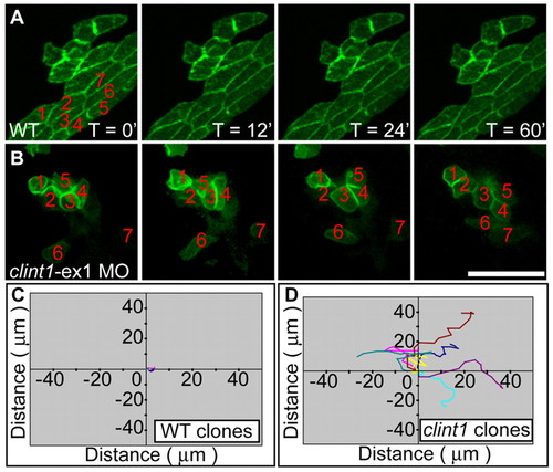
Mesenchymal behavior of epidermal cells in clint1 mutants. (A,B) Still images from in vivo time-lapse microscopy of keratinocyte clusters from uninjected (A) (see Movie 5 in the supplementary material) or clint1-ex1 MO-injected (B) (see Movie 6 in the supplementary material) Tg(β-actin:hras-eGFP) zebrafish embryos that were transplanted into ventral ectoderm of non-transgenic wild-type (A) or clint1 morphant (B) embryos, respectively. Epidermal cells from clint1 morphants exhibit loss of cell-cell contacts and increased motility (B,D). Note the change in position of the numbered cells in B. (C,D) Quantification of migration of the numbered GFP-positive keratinocytes in unlabeled wild type (C) (see Movie 5 in the supplementary material) or clint1-morphant recipients (D) (see Movie 6 in the supplementary material). Scale bar: 50 μm. |