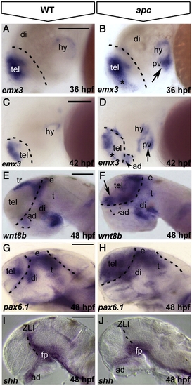
Loss of Apc function leads to mispatterning of the forebrain. Lateral view, anterior to the left. (A?D) As compared to wild-type embryos (A), emx3 expression expands into ventral telencephalon (asterisk) and posterior ventral hypothalamus (arrow) in apc mutants at 36 hpf (B). At 42 hpf (C, D), emx3 further expands into anterior dorsal hypothalamus (dashed line and arrowhead in panel D) and the telencephalic?diencephalic boundary. (E?H) wnt8b (E, F) and pax6.1 (G, H) expand rostrally into the telencephalon and caudally into the thalamus of apc mutants (F, H) at 48 hpf. Dashed line in panels A?H indicates the telencephalic?diencephalic boundary. An additional line in panels G, H indicates the ventral/dorsal midbrain boundary. (I, J) In apc mutants, shh expression is absent from the ZLI and anterior dorsal hypothalamus at 48 hpf (J). Dashed line indicates the ZLI. ad, anterior dorsal hypothalamus; di, diencephalon, e, epithalamus; fp, floorplate; pv, posterior ventral hypothalamus; t, thalamus; ZLI, zona limitans intrathalamica. Scale bar 125 μm.
|
