
Fig. 2
- ID
- ZDB-FIG-090710-10
- Publication
- Martin et al., 2009 - Analysis of heart valve development in larval zebrafish
- Other Figures
- All Figure Page
- Back to All Figure Page
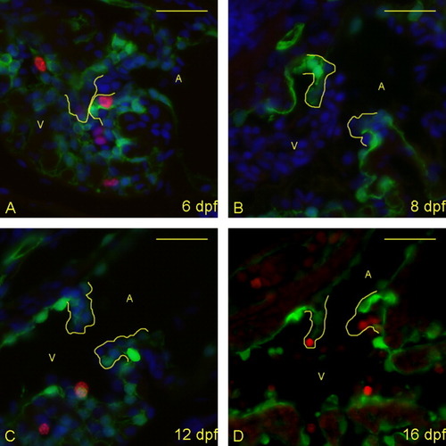
Proliferative cells are seen in the atrioventricular (AV) boundary at 6 and 16 days postfertilization (dpf), but not between these times. A-D: Five micrometer paraffin sections of flk1::GFP transgene-carrying zebrafish embryos/larvae were imaged to identify proliferating cells in the AV boundary and intra-luminal structure. Atria (A) and ventricles (V) are labeled (yellow letters), and yellow lines outline the extent of the AV structure (cushion and/or valve). Staining identifies cell nuclei (DAPI; blue), proliferative cells (anti-bromodeoxyuridine [BrdU]; red), and endothelial cells (anti-GFP; green). (DAPI not shown in panel D to emphasize BrdU-positive cells). Proliferative cells are seen in the developing valves at 6 dpf (A) and 16 dpf (D), but not 8 and 12 dpf (B,C). Scale bar = 20 μm. |