
Fig. 8
- ID
- ZDB-FIG-090504-66
- Publication
- Higashijima et al., 1997 - Mindin/F-spondin family: novel ECM proteins expressed in the zebrafish embryonic axis
- Other Figures
- All Figure Page
- Back to All Figure Page
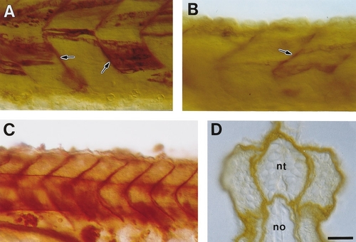
Localization of ectopically expressed Mindin1 protein. All embryos are at about 24 h of development. (A) Lateral view of the zα-actin? mindin1-myc-injected embryo stained with the anti-myc antibody. (B) Lateral view of the zα-actin?mindin1-injected embryo stained with one of the anti-Mindin1 antibodies (Ab-mdn1-6). The antibody may recognize Mindin2 (see Materials and Methods). However, the staining pattern was quite likely derived from ectopically expressed Mindin1 in muscle cells, since such staining was absent in uninjected embryos. Arrows in A and B indicate intense immunoreactivity in the basal lamina. (C) Lateral view of the sCMV?mindin1- myc-injected embryo stained with the anti-myc antibody. (D) Cross section of the sCMV?mindin1-myc-injected embryo stained with the anti-myc antibody. nt, neural tube; no, notochord. Scale bar, 27 μm in A and B, 40 μm in C, and 15 μm in D. |
Reprinted from Developmental Biology, 192, Higashijima, S., Nose, A., Eguchi, G., Hotta, Y., and Okamoto, H., Mindin/F-spondin family: novel ECM proteins expressed in the zebrafish embryonic axis, 211-227, Copyright (1997) with permission from Elsevier. Full text @ Dev. Biol.