
Fig. 7
- ID
- ZDB-FIG-090504-49
- Publication
- Glasgow et al., 1997 - Neuronal and neuroendocrine expression of lim3, a LIM class homeobox gene, is altered in mutant zebrafish with axial signaling defects
- Other Figures
- All Figure Page
- Back to All Figure Page
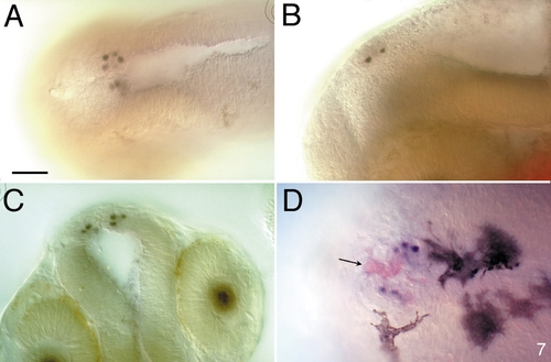
Expression of Lim3 in the epiphysis is restricted to a subset of projection neurons. (A–C) Four to five Lim3-expressing cells are seen on each side of the midline in a ventrolateral position in the epiphysis of a 28-h embryo. (A) Dorsal view. (B) Lateral view. (C) Anterior view. This optical section through the epiphysis, with the diencephalon in focus, is slightly deeper than in Fig. 5D. (D) Dorsal view of a 2-day-old embryo. Whole-mount embryos were double labeled with anti-S-antigen (red) and anti-Lim3 (blue, nuclei). Four to five projection neurons express Lim3 protein. Several cells, especially in the center of the epiphysis, express the photoreceptor marker, S-antigen (arrow). By this stage several prominent melanocytes are seen. Scale bar is 50 μm. |
| Gene: | |
|---|---|
| Antibody: | |
| Fish: | |
| Anatomical Term: | |
| Stage Range: | Prim-5 to Long-pec |
Reprinted from Developmental Biology, 192, Glasgow, E., Karavanov, A.A., and Dawid, I.B., Neuronal and neuroendocrine expression of lim3, a LIM class homeobox gene, is altered in mutant zebrafish with axial signaling defects, 405-419, Copyright (1997) with permission from Elsevier. Full text @ Dev. Biol.