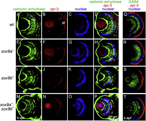
Fluorescent antibody staining of retinal cell types in wild-type and sox9 single and double mutant larvae. (A?P) 5 dpf and (Q?T) 4 dpf. Eye morphology in sox9a mutants was generally similar to that in wild-type siblings. In contrast, sox9b mutants and sox9a;sox9b double mutants had smaller eyes and eye morphology was less organized than in wild-type siblings and sox9a mutants. (A, E, I, M) Retinal Müller glial cells labeled by anti-carbonic anhydrase showed that sox9b mutants (I) and sox9a;sox9b double mutants (M) had fewer Müller glial cells compared to wild-types (A) and sox9a (E) siblings. (B, F, J, N) Photoreceptor cells labeled by zpr-3 showed that fewer and thinner photoreceptor cells were present in sox9b mutants (J) and sox9a;sox9b double mutants (N) compared to wild-type (B) and sox9a (F) siblings. (C, G, K, O) Nuclear layers of the retina stained by nuclear dye (TO-PRO-3 iodide) showed that all the nuclear layers were present in sox9 mutants. (D, H, L, P) green: anti-carbonic anhydrase, red: zpr-3, and blue: nuclear staining. (Q?T) Retinal amacrine cells labeled by anti-GABA: green, photoreceptor cell labeled by zpr-3: red, and nuclear layers of the retina staining by nuclear dye (TO-PRO-3 iodide): blue. The results showed that sox9a mutants (R), sox9b mutants (S) and sox9a;sox9b double mutants (T) have fewer organized amacrine cells and photoreceptor cells compared to wild-type siblings (Q). Scale bar: 100 μm.
|
