
|
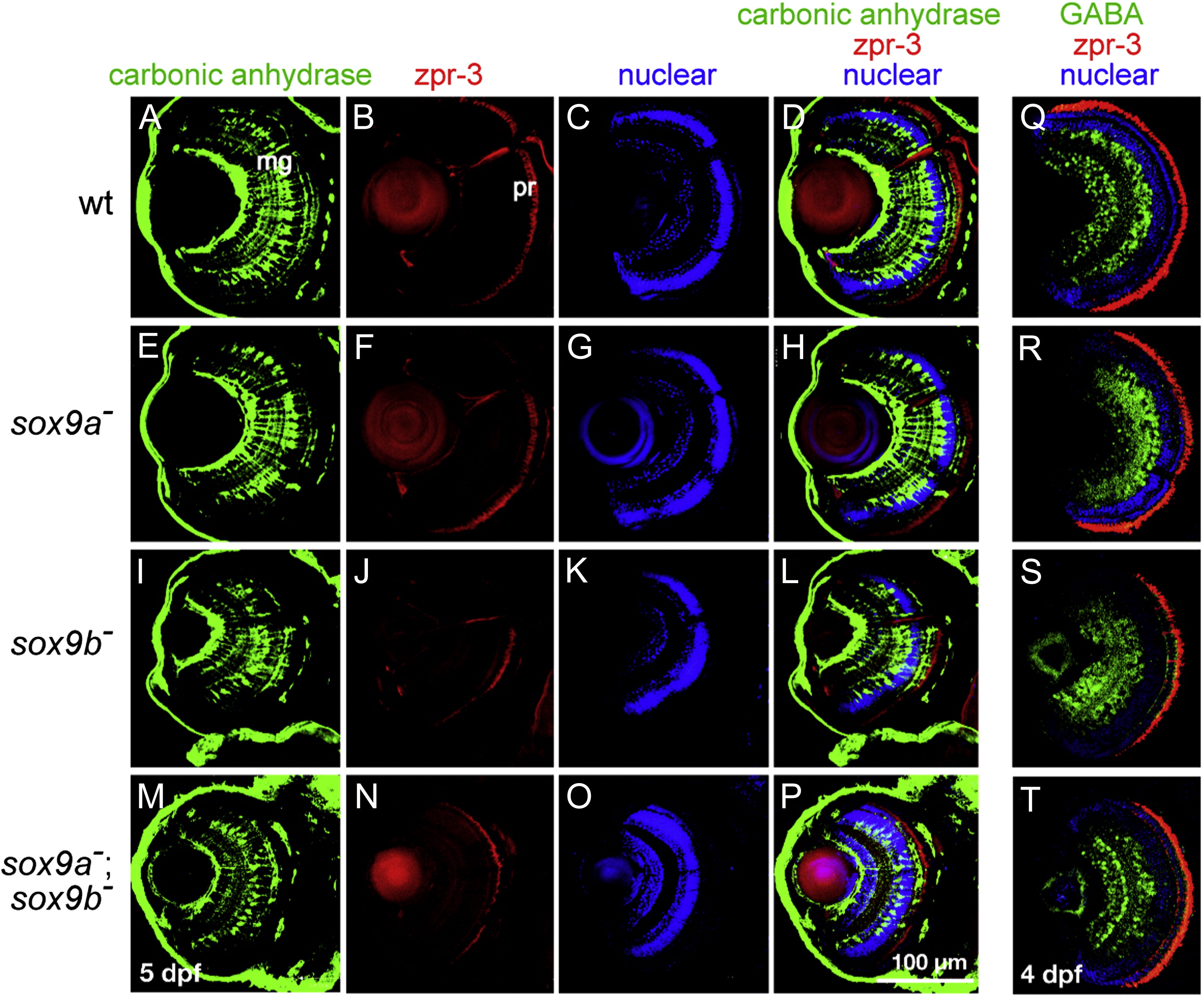
Fig. 6 Fluorescent antibody staining of retinal cell types in wild-type and sox9 single and double mutant larvae. (A?P) 5 dpf and (Q?T) 4 dpf. Eye morphology in sox9a mutants was generally similar to that in wild-type siblings. In contrast, sox9b mutants and sox9a;sox9b double mutants had smaller eyes and eye morphology was less organized than in wild-type siblings and sox9a mutants. (A, E, I, M) Retinal Müller glial cells labeled by anti-carbonic anhydrase showed that sox9b mutants (I) and sox9a;sox9b double mutants (M) had fewer Müller glial cells compared to wild-types (A) and sox9a (E) siblings. (B, F, J, N) Photoreceptor cells labeled by zpr-3 showed that fewer and thinner photoreceptor cells were present in sox9b mutants (J) and sox9a;sox9b double mutants (N) compared to wild-type (B) and sox9a (F) siblings. (C, G, K, O) Nuclear layers of the retina stained by nuclear dye (TO-PRO-3 iodide) showed that all the nuclear layers were present in sox9 mutants. (D, H, L, P) green: anti-carbonic anhydrase, red: zpr-3, and blue: nuclear staining. (Q?T) Retinal amacrine cells labeled by anti-GABA: green, photoreceptor cell labeled by zpr-3: red, and nuclear layers of the retina staining by nuclear dye (TO-PRO-3 iodide): blue. The results showed that sox9a mutants (R), sox9b mutants (S) and sox9a;sox9b double mutants (T) have fewer organized amacrine cells and photoreceptor cells compared to wild-type siblings (Q). Scale bar: 100 μm.
Reprinted from Developmental Biology, 329(1), Yokoi, H., Yan, Y.L., Miller, M.R., Bremiller, R.A., Catchen, J.M., Johnson, E.A., and Postlethwait, J.H., Expression profiling of zebrafish sox9 mutants reveals that Sox9 is required for retinal differentiation, 1-15, Copyright (2009) with permission from Elsevier. Full text @ Dev. Biol.