
Fig. 3
- ID
- ZDB-FIG-090320-38
- Publication
- Sims Jr et al., 2009 - Connexin43 regulates joint location in zebrafish fins
- Other Figures
- All Figure Page
- Back to All Figure Page
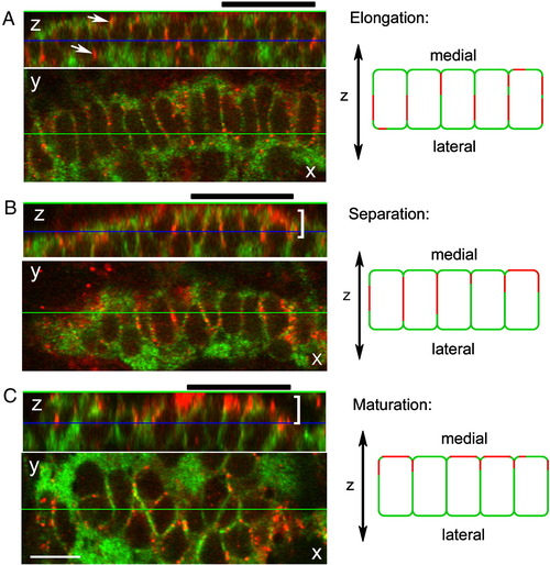
Cx43 localization during joint morphogenesis. A single image from a Z-stack collected by confocal microscopy is shown in the X?Y plane. The display on top represents a slice through the Z-stack along the horizontal green line (i.e., the X?Z plane). The blue line in this display indicates the location of the front image within the Z-stack. During elongation (A), Cx43 (arrows) is found surrounding the ZNS5 positive cells. During separation (B) and maturation (C), Cx43 appears polarized (brackets) toward the medial face of the ZNS5-positive cells. A group of five cells from the X?Z planes are represented by cartoons to the right of each image. Thick black bars over each X?Z plane identify the cells represented by cartoons at the right. Scale bar, 10 μm. |
| Antibodies: | |
|---|---|
| Fish: | |
| Anatomical Term: | |
| Stage: | Adult |
Reprinted from Developmental Biology, 327(2), Sims Jr, K., Eble, D.M., and Iovine, M.K., Connexin43 regulates joint location in zebrafish fins, 410-418, Copyright (2009) with permission from Elsevier. Full text @ Dev. Biol.