|
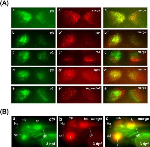
Double fluorescence in situ hybridizations of habenula-expressing markers, gfp and sncga in Tg(sncga:GFP) fish. A: Expression domains of sncga (a′), lov (b′), ron (c′), cpd2 (d′) and f-spondin2 (e′) along with gfp (a-e) in habenula in Tg(sncga:GFP) fish at 3 days postfertilization (dpf). a″-e″, merged image of a and a′, b and b′, c and c′; d and d′, and e and e′, respectively. B: Double fluorescence in situ hybridization of sncga and gfp in an Tg(sncga:GFP) fish at 3 dpf. Panel a shows the staining of gfp mRNA, whereas panel b shows the staining of sncga mRNA. Panel c shows the merged image, in which gfp and sncga staining completely overlapped. gcl, retinal ganglion cell layer; GFP, green fluorescent protein; gP, posterior lateral line ganglion; Ha, habenula; hb, hindbrain; l, lens; mb, midbrain.
|