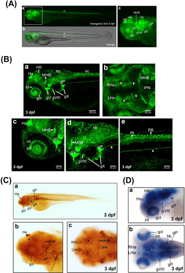
The expression pattern of green fluorescent protein (GFP) in transgenic zebrafish Tg(sncga:GFP) line. Microinjection of the expression construct into zebrafish embryos at the one- to two-cell stage and generation of transgenic GFP line by means of Tol2-mediated transgenesis were described in the text. A: Images from Tg(sncga:GFP) transgenic line at 3 days postfertilization (dpf). Images of bright field and fluorescence are merged and shown in panel b, while fluorescence images are shown in panel a. sc, spinal cord; nc, notochord. B: Confocal image analysis. Zebrafish larva of transgenic Tg(sncga:GFP) line at 3 dpf were collected and fixed with 4% paraformaldehyde. After treatment of FocusClear, embryos were subjected to high-resolution confocal image analysis. All embryos are shown with the anterior to the left. Embryos in a, c, d, e are shown in lateral view, while embryos in b are shown in dorsal view. White arrowheads indicated habenulo-interpeduncular projection (panel b) and posterior lateral line projection (panels d, e). C: Whole-mount immunostaining of Tg(sncga:GFP) embryos by polyclonal antibody against GFP. Embryos in a and b are shown in lateral view, while embryos in c are shown in dorsal view. The habenulo-interpeduncular projection was labeled by black arrowheads. D: Expression patterns of sncga mRNA in zebrafish embryo at 3 dpf. gcl, retinal ganglion cell layer; gP, posterior lateral line ganglion; gV, trigeminal ganglion; gVIII, statoacoustic ganglion; gX, vagal ganglion; Ha, habenula; hb, hindbrain; mb, midbrain; IN, interneuron; inl, inner nuclear layer; IPN, interpeduncular nucleus; l, lens; LHa, left-side habenula; MHB, midbrain-hindbrain boundary; RB, Rohon-Beard neuron; RHa, right-side habenula.
|
