
Fig. 6
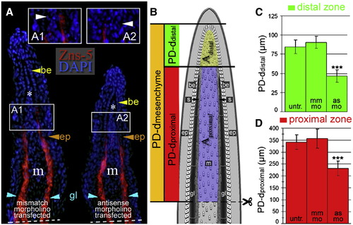
smp knock-down alters morphological proportions in regenerating fin rays. (A) Immunohistochemical labeling of lepidotrichial scleroblasts (red) with the Zns-5 antibody on coronal sections through a regenerating caudal fin from mismatch- and antisense-morpholino-transfected fins 3 dpa. Nuclei were stained with DAPI (blue). Inset panels (A1 and A2) show the most distal end of the Zns-5 labeling: the boundary between distal and proximal zones. Dashed lines show amputation planes. (B) A diagram of a regenerating fin illustrating different regions based on the presence of scleroblasts (s) and Zns-5. (C and D) The graph of the direct measurements of the distal (scleroblast negative) compartment (C) and proximal (scleroblast positive) compartment (D) along the proximodistal axis (PD) between untransfected, mismatch-transfected and antisense-morpholino-transfected fins. Thirty-two cryosections for Zns-5 IHC were measured. (***: p < 0.001, Student's t-test). |
| Antibody: | |
|---|---|
| Fish: | |
| Condition: | |
| Knockdown Reagent: | |
| Anatomical Term: | |
| Stage: | Adult |
Reprinted from Developmental Biology, 325(2), Kizil, C., Otto, G.W., Geisler, R., N�sslein-Volhard, C., and Antos, C.L., Simplet controls cell proliferation and gene transcription during zebrafish caudal fin regeneration, 329-340, Copyright (2009) with permission from Elsevier. Full text @ Dev. Biol.