
Fig. 6
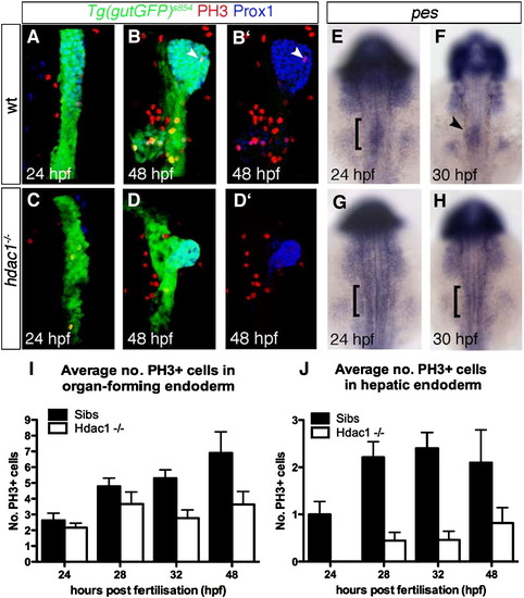
Loss of hdac1 results in reduced cell proliferation in the organ-forming endoderm. (A?D′) Ventral projections using Tg(gutGFP)s854 line, stained for PH3-positive cells (red) and Prox1-positive hepatoblasts (blue) at 24 hpf (A, C), and 48 hpf (B, D); dorsal to the top. (E?H) In situ hybridisation analysis of pes expression. Wild type embryos express pes in the organ-forming region at 24 hpf (bracket, E) and hepatic region at 30 hpf (arrowhead, F). hdac1 mutants lack pes expression in the organ-forming endoderm at both 24 and 30 hpf (brackets, G, H). (I, J) hdac1 mutants display a decreased number of PH3-positive cells the endoderm and liver between 24 and 48 hpf. Numbers are supplied in Table 1 and Table 2. |
| Genes: | |
|---|---|
| Antibody: | |
| Fish: | |
| Anatomical Terms: | |
| Stage Range: | Prim-5 to Long-pec |
Reprinted from Developmental Biology, 322(2), Noėl, E.S., Casal-Sueiro, A., Busch-Nentwich, E., Verkade, H., Dong, P.D., Stemple, D.L., and Ober, E.A., Organ-specific requirements for Hdac1 in liver and pancreas formation, 237-250, Copyright (2008) with permission from Elsevier. Full text @ Dev. Biol.