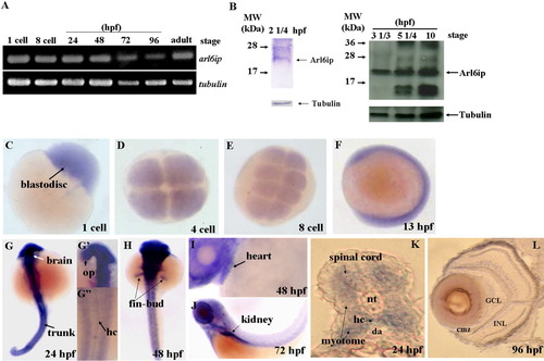
Detection of the existence of arl6ip transcript and protein in zebrafish embryos using RT-PCR (A) and Western blot (B), respectively. The spatial expression of arl6ip mRNA was detected by whole-mount in situ hybridization (C-L). The arl6ip mRNA was first detected in blastodisc at the 1-cell stage (A,C), suggesting that the arl6ip transcript is maternally inherited. RT-PCR revealed that the arl6ip transcript was also detected at the 8-cell stage, 24, 48, 72, 96 hpf, and adulthood, when tubulin mRNA was used as a positive control (A). Total proteins were extracted from the wild-type embryos at 22 1/2, 3 1/3, 5 1/4 or 10 hpf, and the rabbit polyclonal antibody against ARMER was used to perform Western blot analysis when endogenous β-Tubulin was used as a control (B). Arl6ip was present at 2 1/4, 3 1/3, 5 1/4, and 10 hpf (indicated by arrow). Whole-mount in situ hybridization showed that the arl6ip was expressed ubiquitously in developing embryos at the 1-, 4-, and 8-cell stages and 13 hpf (C-F). The arl6ip was displayed in the brain, optic primordia (op), spinal cord, myotome, hypochord (hc), trunk, fin-bud, heart, kidney, and retina from 24 to 96 hpf (G-L). Dorsal view of arl6ip staining (G-G″,H). Lateral view of arl6ip expressions (F,I,J). arl6ip was expressed in retina by cross-sectioning of the eyes at 96 hpf (L). nt: notochord; da: dorsal aorta; GCL: ganglion cell layer; INL: inner nuclear layer; cmz: ciliary marginal zone; L: lens; hpf: hours post-fertilization of zebrafish embryos.
|
