FIGURE

Fig. S1

ID
ZDB-FIG-081219-20
Publication
Pistocchi et al., 2008 - Identification and expression pattern of zebrafish prox2 during embryonic development
Other Figures
All Figure Page
Back to All Figure Page
Fig. S1

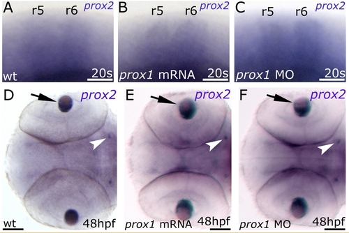
prox1 is not required for proper prox2 expression. (A,B,C) Lateral view. (D,E,F) Dorsal view. Anterior is left in all panels. (A) 20 s control embryo. (B) 20 s embryo injected with prox1 mRNA. (C) 20 s embryo injected with prox1MO. (D) 48 hpf control embryo. (E) 48 hpf embryo injected with prox1 mRNA. (F) 48 hpf embryo injected with prox1MO. prox2 expression was not altered in the rhombomeres (r5 and r6), anterior cranial ganglia (white arrowheads) and lens (arrows) in any of the conditions analyzed. The following abbreviations are used: r, rhombomeres; MO, morpholino oligonucleotide. Scale bars indicate 20 μm (A,B,C) and 25 μm (D,E,F).

Expression Data
Gene:
Fish:
Knockdown Reagent:
Anatomical Terms:
Stage Range: 20-25 somites to Long-pec

Expression Detail
Antibody Labeling
Phenotype Data

Phenotype Detail
Acknowledgments
This image is the copyrighted work of the attributed author or publisher, and ZFIN has permission only to display this image to its users. Additional permissions should be obtained from the applicable author or publisher of the image. Full text @ Dev. Dyn.