
FIGURE
Fig. 4
- ID
- ZDB-FIG-081117-29
- Publication
- Gutzman et al., 2008 - Formation of the zebrafish midbrain-hindbrain boundary constriction requires laminin-dependent basal constriction
- Other Figures
- All Figure Page
- Back to All Figure Page
Fig. 4
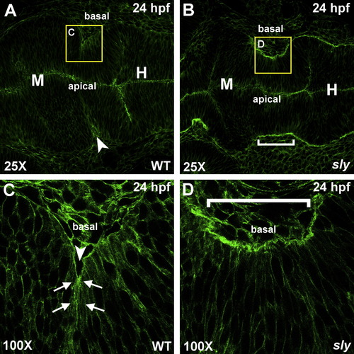
Actin is localized basally at the MHBC in wild type embryos. (A–D) Phalloidin stained wild type (A and C) and sly (B and D) embryos that were fixed at 24 hpf. Stained embryos were flat mounted in glycerol and imaged by confocal microscopy at 25X (A and B) and 100X (C and D). Arrows indicate points of basal actin accumulation in wild type embryos. Anterior is to the left in all images. Arrowhead indicates the MHBC in wild type and a bracket indicates the MHBC region in sly mutants. M, midbrain; H, hindbrain. |
Expression Data
Expression Detail
Antibody Labeling
Phenotype Data
| Fish: | |
|---|---|
| Observed In: | |
| Stage: | Prim-5 |
Phenotype Detail
Acknowledgments
This image is the copyrighted work of the attributed author or publisher, and
ZFIN has permission only to display this image to its users.
Additional permissions should be obtained from the applicable author or publisher of the image.
Reprinted from Mechanisms of Development, 125(11-12), Gutzman, J.H., Graeden, E.G., Lowery, L.A., Holley, H.S., and Sive, H., Formation of the zebrafish midbrain-hindbrain boundary constriction requires laminin-dependent basal constriction, 974-983, Copyright (2008) with permission from Elsevier. Full text @ Mech. Dev.