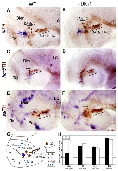
Dkk1 selectively affects DA cell number, but not neighboring basal diencephalic neurons. (A-F) Lateral views of either WT zebrafish embryos (A,C,E) or their dkk1 mRNA-injected siblings (B,D,F). Embryos were fixed at 48 hpf and subjected to whole-mount in situ hybridization with RNA probes directed against the hypothalamic neuropeptide isotocin (zebrafish ortholog of oxytocin) (it; A,B), hypocretin (hcrt; C,D) or somatostatin (ss; E,F). All specimens were then subjected to immunostaining with an anti-tyrosine hydroxylase (TH) antibody to detect DA neurons. (G) Schematic representation of the examined diencephalic cell types in a 48-hpf WT embryo. The positions of DA group 2 (Gr. 2) and groups 3-6 (Gr. 3 to 6) are indicated. (H) Bar chart presenting average counts of dat-expressing DA group 2 cells (DA), isotocin (IT), hypocretin/orexin (Hcrt) or somatostatin (SS) expressing cells in WT and dkk1-injected embryos at 48 hpf. The number of embryos analyzed (n) is shown below. Dien, diencephalon; LC, locus coeruleus; Po, preoptic area; Tel, telencephalon. Scale bar: 50 μm.
|
