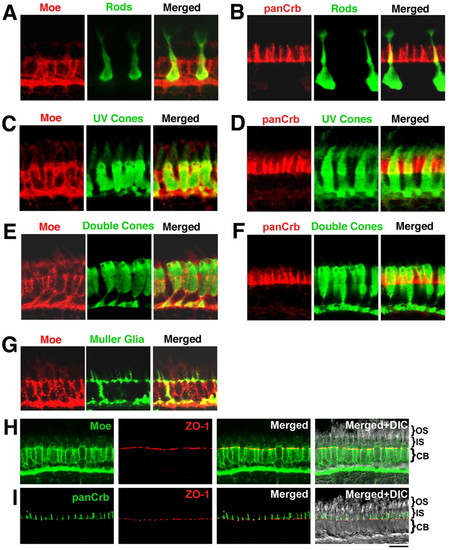
Localization of Moe and panCrb in specific retinal cell types at 5 dpf. (A) Merged guinea pig anti-Moe labeling of rod photoreceptors (in transgenic Xop-GFP fish) with rabbit anti-GFP. (B) Merged rabbit anti-panCrb labeling of GFP+ rod photoreceptors with GFP (in transgenic Xop-GFP fish). (C) Merged guinea pig anti-Moe labeling of UV cones with GFP (in transgenic UV-GFP fish) labeled with mouse anti-GFP. (D) Merged rabbit anti-panCrb labeling of UV cones with GFP (in transgenic UV-GFP fish) labeled with mouse anti-GFP. (E) Merged rabbit anti-Moe labeling of double cones with mouse Zpr-1 antibodies. (F) Merged rabbit anti-panCrb labeling of double cones with mouse Zpr-1 antibodies. (G) Merged guinea pig anti-Moe labeling of Müller glial cells with rabbit anti-Carbonic anhydrase II antibodies. (H) Rabbit anti-Moe, mouse anti-ZO-1, merged anti-Moe and anti-ZO-1, and merged with corresponding DIC image. (I) Rabbit anti-panCrb, mouse anti-ZO-1, merged anti-Moe and anti-ZO-1, and merged with corresponding DIC image. Scale bar: 10 μm. CB, cell bodies.
|
