|
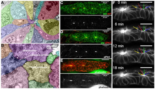
Rosettes are radial clusters of apically constricted epithelial cells. (A) Electron micrographs of an apical section of a rosette; false-colouring highlights individual cells. (B) Close-up of the centre of the rosette, corresponding to the framed area in A. (C-E) Immunostaining using antibodies against GFP and ZO-1 (C), GFP and aPKC (D), and GFP plus phalloidin staining (E). Tight junction protein ZO1 and aPKC are only expressed at the center of each rosette; phalloidin-labeling reveals enriched actin at centre of rosette. (F) Time-lapse analysis of wild-type rosette formation reveals a coordinated constriction of the apical surfaces of several cells. Arrows point to the vertices of these cells as they coalesce. Scale bars: 20 μm (unless stated otherwise).
|