
FIGURE
Fig. 4
- ID
- ZDB-FIG-080702-46
- Publication
- Hyatt et al., 1996 - Retinoic acid alters photoreceptor development in vivo
- Other Figures
- All Figure Page
- Back to All Figure Page
Fig. 4
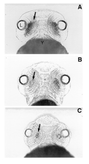
Ventral views of rhodopsin expression in whole mount in situ preparations at day 3 pf in control embryos (A) and embryos treated with the RA-synthetic competitive inhibitor citral (B and C). (A) Rhodopsin expression (arrow) extends across the ventral retina in control embryos at day 3. (B) After treatment with 3 μM citral between days 2 and 3 pf, the level of rhodopsin expression (arrow) is significantly reduced. (C) After treatment with 6 μM citral, rhodopsin expression is further reduced such that only a small number of rods (arrow) are observed within the retina at day 3 pf. L, lens; Y, yolk. |
Expression Data
Expression Detail
Antibody Labeling
Phenotype Data
Phenotype Detail
Acknowledgments
This image is the copyrighted work of the attributed author or publisher, and
ZFIN has permission only to display this image to its users.
Additional permissions should be obtained from the applicable author or publisher of the image.
Full text @ Proc. Natl. Acad. Sci. USA