
|
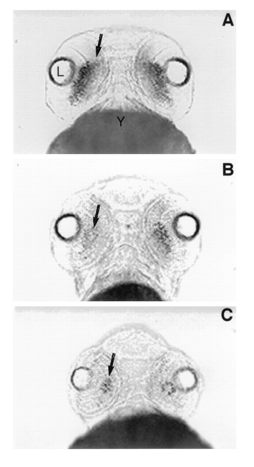
Fig. 4 Ventral views of rhodopsin expression in whole mount in situ preparations at day 3 pf in control embryos (A) and embryos treated with the RA-synthetic competitive inhibitor citral (B and C). (A) Rhodopsin expression (arrow) extends across the ventral retina in control embryos at day 3. (B) After treatment with 3 μM citral between days 2 and 3 pf, the level of rhodopsin expression (arrow) is significantly reduced. (C) After treatment with 6 μM citral, rhodopsin expression is further reduced such that only a small number of rods (arrow) are observed within the retina at day 3 pf. L, lens; Y, yolk.