Fig. 1
- ID
- ZDB-FIG-080326-28
- Publication
- Boldajipour et al., 2008 - Control of chemokine-guided cell migration by ligand sequestration
- Other Figures
- All Figure Page
- Back to All Figure Page
|
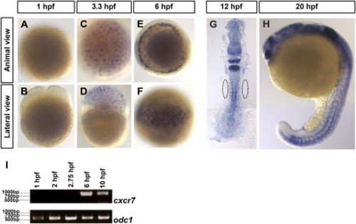
Expression Pattern of cxcr7 (A?H) Distribution of cxcr7 mRNA in wild-type embryos during the first 20 hr of development. In situ hybridization using a cxcr7-specific probe shows no staining in four-cell stage embryos (A and B), weak uniform expression at 3.3 hpf (C and D), and enhanced cxcr7 expression in a ring of deep cells at 6 hpf (E and F). At later stages of development (G and H), uniform cxcr7 expression with enhanced expression in mesoderm derivatives and in the nervous system is detected, but no expression is observed at the region where the PGCs are located (encircled domains). (I) Absence of maternally provided cxcr7 mRNA as determined by RT-PCR at the indicated stages. Control reactions are presented in which primers specific for the maternally provided ornithine decarboxylase1 (odc1) mRNA were used. |
| Genes: | |
|---|---|
| Fish: | |
| Anatomical Terms: | |
| Stage Range: | 4-cell to 20-25 somites |
Reprinted from Cell, 132(3), Boldajipour, B., Mahabaleshwar, H., Kardash, E., Reichman-Fried, M., Blaser, H., Minina, S., Wilson, D., Xu, Q., and Raz, E., Control of chemokine-guided cell migration by ligand sequestration, 463-473, Copyright (2008) with permission from Elsevier. Full text @ Cell

