FIGURE
Fig. 2
- ID
- ZDB-FIG-080326-119
- Publication
- Bonner et al., 2008 - Proliferation and patterning are mediated independently in the dorsal spinal cord downstream of canonical Wnt signaling
- Other Figures
- All Figure Page
- Back to All Figure Page
Fig. 2
|
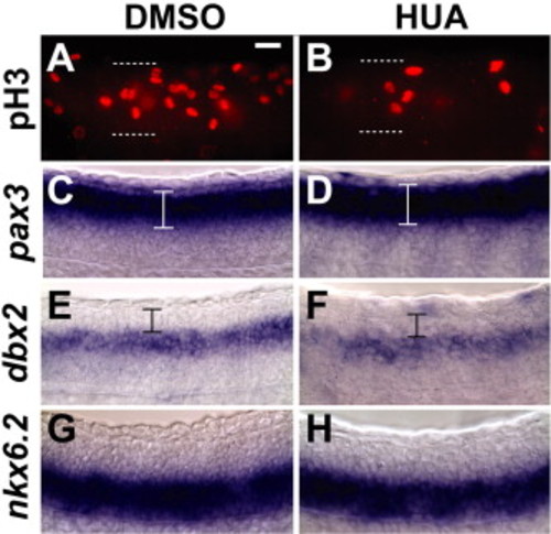
Reduction of cell proliferation does not cause spinal cord patterning defects. (A, B) Anti-phospho histone H3 staining reveals a decrease in proliferative cells in the HUA-treated spinal cord. (C, D) Expression of pax3 is unaffected in embryos treated with HUA for 12 h. (E, F) The dbx2 expression domain is unchanged in HUA embryos although an overall decrease in signal was observed. Expression in the dorsal epidermis is non-specific. (G, H) Expression of nxk6.2 is unaffected in HUA-treated embryos. Lateral mounts are shown. Scale bar = 10 μm. |
Expression Data
| Genes: | |
|---|---|
| Fish: | |
| Condition: | |
| Anatomical Term: | |
| Stage: | Prim-5 |
Expression Detail
Antibody Labeling
Phenotype Data
Phenotype Detail
Acknowledgments
This image is the copyrighted work of the attributed author or publisher, and
ZFIN has permission only to display this image to its users.
Additional permissions should be obtained from the applicable author or publisher of the image.
Reprinted from Developmental Biology, 313(1), Bonner, J., Gribble, S.L., Veien, E.S., Nikolaus, O.B., Weidinger, G., and Dorsky, R.I., Proliferation and patterning are mediated independently in the dorsal spinal cord downstream of canonical Wnt signaling, 398-407, Copyright (2008) with permission from Elsevier. Full text @ Dev. Biol.

