|
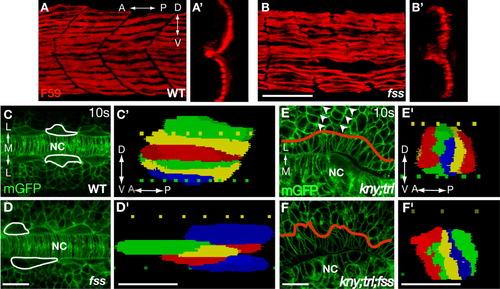
Somitic boundaries are dispensable for the shape changes and lateral migration of the adaxial cells. A, B: Lateral views of embryos stained with F59 antibody. A', B': 3D reconstructed transverse sections of the slow muscle fibers shown in A and B. C-F: Dorsal views of the embryos expressing mGFP. In C and D, white lines outline the selected adaxial cells. In E and F, red lines show the boundary between the adaxial cells and the lateral somitic cells. Arrowheads in E mark the somitic boundaries, which are missing in the kny;tri;fss embryos (F). C'-F': 3D reconstruction of the adaxial cells within the third somite at the 10-somite stage (14 hpf). Lateral views. A, anterior; D, dorsal; L, lateral; M, medial; NC, notochord; P, posterior; V, ventral. Scale bars = 50 μm (A,B, A',B'); 20 μm (C-F, C'-F').
|