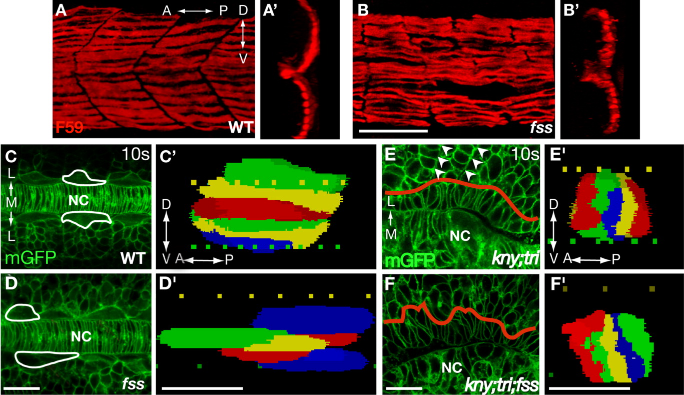
Fig. 4 Somitic boundaries are dispensable for the shape changes and lateral migration of the adaxial cells. A, B: Lateral views of embryos stained with F59 antibody. A', B': 3D reconstructed transverse sections of the slow muscle fibers shown in A and B. C-F: Dorsal views of the embryos expressing mGFP. In C and D, white lines outline the selected adaxial cells. In E and F, red lines show the boundary between the adaxial cells and the lateral somitic cells. Arrowheads in E mark the somitic boundaries, which are missing in the kny;tri;fss embryos (F). C'-F': 3D reconstruction of the adaxial cells within the third somite at the 10-somite stage (14 hpf). Lateral views. A, anterior; D, dorsal; L, lateral; M, medial; NC, notochord; P, posterior; V, ventral. Scale bars = 50 μm (A,B, A',B'); 20 μm (C-F, C'-F').
Image
Figure Caption
Figure Data
Acknowledgments
This image is the copyrighted work of the attributed author or publisher, and
ZFIN has permission only to display this image to its users.
Additional permissions should be obtained from the applicable author or publisher of the image.
Full text @ Dev. Dyn.

