
Fig. 3
- ID
- ZDB-FIG-071017-2
- Publication
- Chong et al., 2001 - Expression pattern of two zebrafish genes, cxcr4a and cxcr4b
- Other Figures
- All Figure Page
- Back to All Figure Page
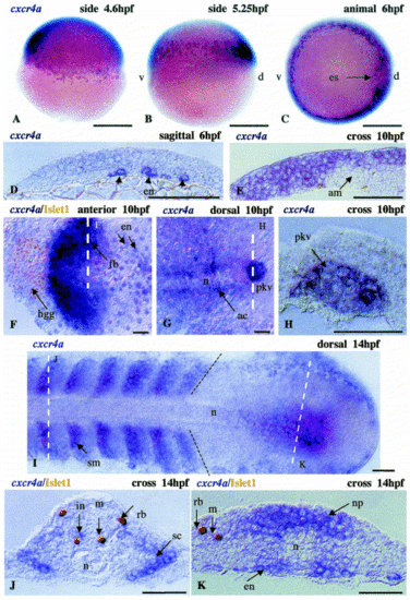
Embryonic expression of cxcr4a. The expression pattern of cxcr4a includes: (A?D), the early endoderm (en); (G,H), the primordium of Kupffer′s vesicle (pkv); and (E,F), the forebrain (fb). (G) The expression appeared relatively late in adaxial cells (ac). (D) is a sagittal section of the embryo shown in (C). In (F), the white broken lines define a section shown in (E). In (G), the white line defines a section shown in (H). During somitogenesis, cxcr4a is expressed in somites (sm) immediately after their separation from the unsegmented lateral mesodermal plate (black dotted line) and in the tail bud (I). In addition, ventral interneurons (in) express this gene (J). In somites, expression of cxcr4a is confined to the sclerotome (sc) (J). In the tail bud (K), cxcr4a expression is found mainly in the neural plate (np) and endoderm (en). am, axial mesoderm; d, dorsal; es, embryonic shield; hgg, hatching gland; n, notochord; rb, Rohon?Beard sensory cells; v, ventral. Black arrowheads in (D) indicate endodermal cells. The broken black line represents the border of segmented and unsegmented lateral mesoderm. Anterior in all figures is oriented to the left unless otherwise indicated. Broken white lines represent the section level. Scale bar in (A?C), 250 μm; remainder, 50 μm. |
| Gene: | |
|---|---|
| Fish: | |
| Anatomical Terms: | |
| Stage Range: | Shield to 10-13 somites |
Reprinted from Mechanisms of Development, 109(2), Chong, S.-W., Emelyanov, A., Gong, Z., and Korzh, V., Expression pattern of two zebrafish genes, cxcr4a and cxcr4b, 347-354, Copyright (2001) with permission from Elsevier. Full text @ Mech. Dev.