
Fig. 7
- ID
- ZDB-FIG-070924-11
- Publication
- Chen et al., 2005 - A unique role for 6-O sulfation modification in zebrafish vascular development
- Other Figures
- All Figure Page
- Back to All Figure Page
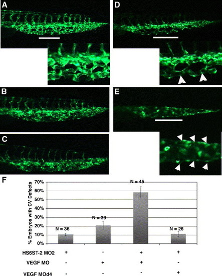
HS6ST-2 and VEGF-A interact during caudal vein plexus formation in vivo. (A) Uninjected Tg(fli-1:EGFP) embryo at 33 hpf under FITC channel, with the inset displaying an enlargement of a segment in the venous plexus. (B) Embryo injected with 0.5 ng of HS6ST-2 MO2 displaying a venous plexus without any obvious defect. (C) Embryo injected with 1 ng of VEGF-A MO, displaying a venous plexus without any obvious defect. (D?E) Embryo co-injected with HS6ST-2 MO2 and VEGF-A MO displaying defective branching morphogenesis of the venous plexus as indicated by the formation of large loops (arrowheads in inset of D) and a single cavernous vessel with no branching (arrowheads in inset of E). The lines in A, D and E indicate regions in the venous plexus that are displayed in the insets. (F) A summary of two independent experiments is shown. Embryos co-injected with HS6ST-2 MO2 (0.5 ng) and VEGF-A MO (1 ng) exhibit a more than additive increase in the frequency (�SEM) of embryos with branching defects in the caudal vein plexus (column 3) compared to embryos injected with HS6ST-2 MO2 or VEGF-A MO only. In contrast, no synergy is observed in the embryos co-injected with HS6ST-2 MO and a four-base mismatch MO (d4, 1 ng) against VEGF-A (column 4). |
Reprinted from Developmental Biology, 284(2), Chen, E., Stringer, S.E., Rusch, M.A., Selleck, S.B., and Ekker, S.C., A unique role for 6-O sulfation modification in zebrafish vascular development, 364-376, Copyright (2005) with permission from Elsevier. Full text @ Dev. Biol.