
Fig. 5
- ID
- ZDB-FIG-050906-9
- Publication
- Chen et al., 2005 - A unique role for 6-O sulfation modification in zebrafish vascular development
- Other Figures
- All Figure Page
- Back to All Figure Page
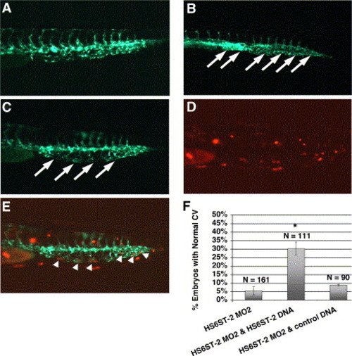
Co-injection of zebrafish HS6ST-2 expression construct alleviates the caudal vein defects in HS6ST-2 morphant embryos. (A) Uninjected Tg(fli-1:EGFP) embryo at 33 hpf under FITC illumination. (B) HS6ST-2 MO2-injected embryo with defective caudal vein branching as indicated by the formation of a cavernous vessel (arrows). (C) Embryo co-injected with HS6ST-2 MO2 and a mixed solution of HS6ST-2 DNA and DsRed DNA, showing the restoration of the venous plexus (arrows) under FITC illumination. DsRed is introduced as a tracer to facilitate the identification of DNA-injected embryos. (D) DsRed expression in the same co-injected embryo showing mosaic expression pattern in the tail under TRITC illumination. (E) Overlay of panels C and D, showing DsRed expressing-cells, correlating with areas in the restored venous plexus (arrowheads). (F) Summary of four independent co-injection experiments. The test DNA (HS6ST-2 expression construct) or the control DNA (DsRed expression construct) was injected into embryos separately injected with HS6ST-2 MO2. The introduction of HS6ST-2 protein by the DNA injection alleviates the caudal vein defects in HS6ST-2 MO2-injected embryos (compare column 2 to column 1). Asterisk (*) denotes statistical significance with P < 0.0005. In contrast, a separate injection of control DNA into HS6ST-2 MO2-injected embryos does not affect the frequency of caudal vein defects in HS6ST-2 MO2-injected embryos (compare column 3 to column 1; P > 0.2). N indicates the total number of embryos scored. ±SEM. |
Reprinted from Developmental Biology, 284(2), Chen, E., Stringer, S.E., Rusch, M.A., Selleck, S.B., and Ekker, S.C., A unique role for 6-O sulfation modification in zebrafish vascular development, 364-376, Copyright (2005) with permission from Elsevier. Full text @ Dev. Biol.