
Fig. 4
- ID
- ZDB-FIG-070821-11
- Publication
- Shi et al., 2005 - Zebrafish pitx3 is necessary for normal lens and retinal development
- Other Figures
- All Figure Page
- Back to All Figure Page
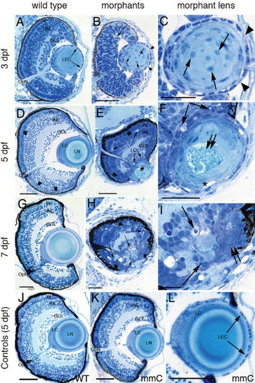
Histological analysis of the morphant lens and retina phenotypes. Stained eye sections of wild type (left column) and anti-pitx3 morphants at low and high magnification (center and right columns, respectively) at 3, 5 and 7 dpf. Sections of control eyes (wild type and mismatch control) are shown in the bottom row of panels (J–L). Retinal cell pyknosis (long arrows in B, E, F, H and I) and retention of the lens fiber cell nuclei (short arrows in B and C) are visible in the morphants, but absent in the controls. At 5 dpf, the epithelial cells in the morphant lens are irregularly shaped and overlie one another in the lens cortex (E, F). The lens epithelial cell layer contains gaps that lack nuclei or exhibits overlapping cells (B and C, arrowheads). Gaps exist between the lens epithelial cells and the underlying fiber cells in the distal lens (E and F, asterisks). Central lens fiber degeneration (double arrows) is observed in the morphant lens (E, F) at 5 dpf, but not in the wild-type lens (D). At 7 dpf, the retinal regression and lens degeneration in the morphant (H, I) are severe relative to wild type (G). The lens and retina of the mismatch control (K, L) appear identical to the wild type (J). Abbreviations: PL, photoreceptor layer; INL, inner nuclear layer; GCL, ganglion cell layer; OpN, optic nerve; LEC, lens epithelial cells; LN, lens nucleus; LC, lens cortex; IPL, inner plexiform layer. The scale bars represent 50 μm. |
Reprinted from Mechanisms of Development, 122(4), Shi, X., Bosenko, D.V., Zinkevich, N.S., Foley, S., Hyde, D.R., Semina, E.V., and Vihtelic, T.S., Zebrafish pitx3 is necessary for normal lens and retinal development, 513-527, Copyright (2005) with permission from Elsevier. Full text @ Mech. Dev.