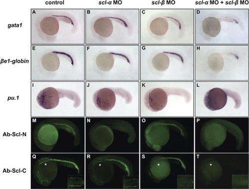
scl-α and -β Are Functionally Redundant in the Initiation of Primitive Hematopoiesis (A?L) WISH of gata1 (A?D), βe1-globin (E?H), and pu.1 (I?L). Similar expression of gata1, βe1-globin, and pu.1 were detected in 20-hpf control embryos (A, E, and I), scl-α morphants (B, F, and J), and scl-β morphants (C, G, and K). In contrast, expression of gata1, βe1-globin, and pu.1 were significantly reduced or absent in scl-α/β co-injected morphants (D, H, and L). All embryos are in lateral view with anterior to the left. (M?T) Immunohistochemistry staining of endogenous Scl proteins in 20-hpf control embryos (M and Q) and scl-α (N and R), scl-β (O and S), and scl-α/β co-injected (P and T) morphants using Ab-Scl-N (M?P) and Ab-Scl-C (Q?T). In scl-α morphants, the Ab-Scl-N (N), but not Ab-Scl-C (R), staining in the ICM was absent, showing that scl-α MO specifically blocks the translation of scl-α. In the scl-β morphants, the Ab-Scl-C staining was selectively abolished in the APLM, where only scl-β is transcribed ([S] asterisk, inset), demonstrating that scl-β MO specifically blocks the protein expression of scl-β. In the scl-α/β co-injected morphants, neither Ab-Scl-N nor Ab-Scl-C staining was detected (P and T). Insets in (Q?T) are dorsal views of the magnified APLM marked by asterisks. Embryos are in lateral view with anterior to the left.
|

