|
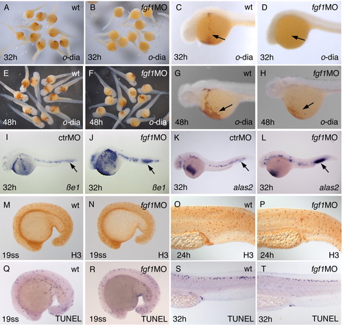
Erythrocyte differentiation is delayed in fgf1 morphants, but cell proliferation and cell death appears to be normal. Labeling of panels is as in Figure 2>. A-H:o-dianisidine staining is missing at 32 hpf in fgf1 morphants (arrows in C,D), but is present at 48 hpf, albeit at reduced levels (arrows in G,H). I-L: The differentiation markers βe1 and alas2 are continuously expressed in fgf1 morphants (arrows point to the posterior intermediate cell mass (ICM), highlighting the accumulation of extra cells in fgf1 morphants). M-P: Immunohistochemistry with an antibody detecting phosphorylated histone-3 (H3) detects mitotic cells. No increased cell proliferation can be detected in fgf1 morphants. Q-T: terminal deoxynucleotidyl transferase-mediated deoxyuridinetriphosphate nick end-labeling (TUNEL) assay detects cell death. No increased cell death can be observed in fgf1 morphants. H3, antibody staining with anti-phosphorylated histone.
|