
Fig. 8
- ID
- ZDB-FIG-060808-244
- Publication
- Stigloher et al., 2006 - Segregation of telencephalic and eye-field identities inside the zebrafish forebrain territory is controlled by Rx3
- Other Figures
- All Figure Page
- Back to All Figure Page
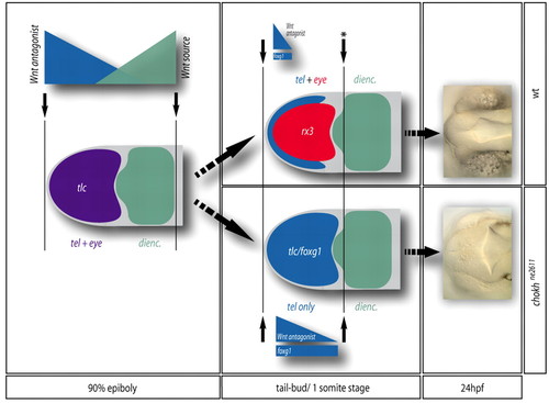
Model for the segregation of the telencephalic and eye anlage during zebrafish forebrain patterning. Patterning boundaries are indicated by black arrows. During gastrulation (left panel), a prevalent view is that the forebrain is patterned as a whole by the opposite activities of a posterior Wnt source located at the level of the posterior diencephalon (green gradient) and Wnt antagonists (purple gradient) located at the ANB (Wilson and Houart, 2004). `High Wnt' defines the presumptive diencephalon (green), whereas `low Wnt' defines an anterior domain (purple) including the presumptive telencephalon and eyes. At the tail-bud stage (middle panel), a posterior patterning boundary (black arrow with star) is set-up in front of the diencephalon, isolating the anterior forebrain. Within the latter domain, Rx3 activity (red) represses tlc expression (blue gradient) and foxg1 (blue bar) and attributes retinal fate to its expressing cells at the expense of a telencephalic fate, thereby segregating retinal and telencephalic identities (top panel). In the absence of Rx3, retinal precursors acquire a telencephalic fate (bottom panel), leading at 24 hpf to the absence of eyes and an enlarged telencephalon (right panel). |