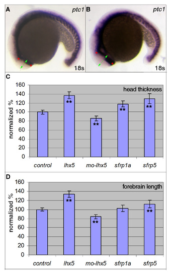
Lhx5, Sfrp1a and Sfrp5 regulate the size of the forebrain. Quantitative measurement of forebrain sizes. (A,B) An uninjected control embryo (A) and an lhx5 mRNA-injected embryo (B) are shown to illustrate how quantitative measurements are carried out. Injected embryos and uninjected control embryos were fixed and processed for in situ hybridization with ptc1 probes. Images of labeled embryos were analyzed with Axiovision software (Carl Zeiss). Two parameters were measured for each image. Green double arrow (head thickness) crosses the apex of forebrain and is approximately perpendicular to the tangent plane at the apex. Forebrain length was defined as the distance between the anterior limit and the diencephalic bending point of ptc1 labeling (red stars). The ptc1 bending point is located at the zona limitans intrathalamica that separates the anterior diencephalon and the posterior diencephalon. (C,D) Measurements were normalized by dividing by the values of uninjected controls and multiplying by 100. ** denotes significant differences between uninjected control and affected injected embryos (t-test, P<0.01). For each measurement, the sample size was n=10.
|

