
Fig. 7
- ID
- ZDB-FIG-060607-5
- Publication
- Ho et al., 2006 - Zebrafish fat-free is required for intestinal lipid absorption and Golgi apparatus structure
- Other Figures
- All Figure Page
- Back to All Figure Page
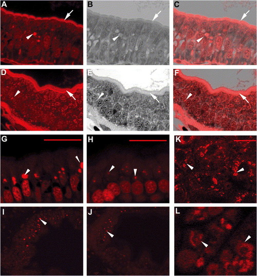
ffr mutants exhibit altered vesicular trafficking and APOA1 targeting A–F) Histological cross-sections through the intestine of 5 dpf ffr (A–C) and wild-type (D–F) larvae. Red fluorescence shows AM1-43 accumulation in plasma membrane (arrows) and intracellular vesicles (arrow head) in ffr mutant and wild-type enterocytes. (B and E) Identical sections in (A) and (D) stained with 1% methylene blue in 1% sodium borate and 1% Azure B (1:1). (C and F) Composites of panels (A) and (B), and (D) and (E), respectively. Note accumulation of AM1-43 in the region of the Golgi of ffr enteroctyes (C). This is best appreciated in histological cross-sections through the intestine of 5 dpf ffr and wild-type larvae following AM1-43 ingestion and cyclodextran wash to remove noninternalized dye within the plasma membrane (G–J). (K–L) Following ffr “knockdown” by MO-1, the apoA1-mRFP fusion protein had a diffuse intracellular distribution (arrowhead in [L]), whereas in control embryos the apoA1-mRFP fusion protein was present within vesicles surrounding the nucleus (arrowhead). The scale bar represents 15 μm. |
| Fish: | |
|---|---|
| Knockdown Reagent: | |
| Observed In: | |
| Stage: | Day 5 |
Reprinted from Cell Metabolism, 3(4), Ho, S.Y., Lorent, K., Pack, M., and Farber, S.A., Zebrafish fat-free is required for intestinal lipid absorption and Golgi apparatus structure, 289-300, Copyright (2006) with permission from Elsevier. Full text @ Cell Metab.