
Fig. 3
- ID
- ZDB-FIG-060410-17
- Publication
- Hammond et al., 2006 - The developing lamprey ear closely resembles the zebrafish otic vesicle: otx1 expression can account for all major patterning differences
- Other Figures
- All Figure Page
- Back to All Figure Page
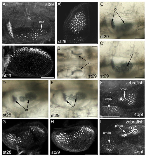
Morphology of stage 28 and 29 L. fluviatilis ears, and comparison with zebrafish. (A-E,G,H) L. fluviatilis; (F,I) zebrafish, focussed medially to reveal maculae. amac, anterior macula; pmac, posterior macula (A-A'',F-I) Projections of confocal z-stacks of FITC-phalloidin-stained ears revealing the actin-rich stereociliary bundles of the sensory hair cells (hcs). (B-E) DIC images of ears in the live lamprey larva. ot, otoconia. (A,C,C',F) Lateral views, anterior to left, dorsal to top; (C,C') different focal planes of the same specimen. (A') Anterior view, dorsal to top, lateral to left. (A'',B) Dorsal views, anterior to left; medial to top in A''; both ears shown in B. (D,E,G-I) Dorsolateral views showing the join region between anterior and posterior areas of the macula; anterior approximately left. Scale bars: 50 μm. |