
Fig. 6
- ID
- ZDB-FIG-060307-7
- Publication
- Sakai et al., 2006 - Semaphorin 3d guides laterality of retinal ganglion cell projections in zebrafish
- Other Figures
- All Figure Page
- Back to All Figure Page
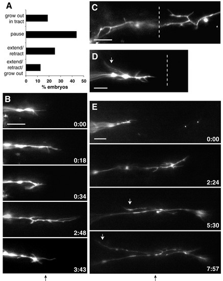
RGC axons show ipsilateral and interstitial projections and reduced growth beyond the midline following Sema3d knockdown. (A) Summary of growth cone behaviors after reaching the chiasm midline in Sema3d morphant embryos. (B) Frames taken from a timelapse movie of a Sema3d morphant embryo show repeated growth cone extensions and retractions at the midline. (C,D) Sema3d knockdown induced interstitial processes, including ipsilateral projections (white arrow). (E) Some processes developed into ipsilateral growth cones (white arrows). Times are indicated in hours:minutes; midlines are indicated by dotted lines or black arrows below frame sequences. Anterior is up in all frames. Scale bars: 10 μm. |