
Fig. 1
- ID
- ZDB-FIG-060301-22
- Publication
- Sato-Maeda et al., 2006 - Sema3a1 guides spinal motor axons in a cell- and stage-specific manner in zebrafish
- Other Figures
- All Figure Page
- Back to All Figure Page
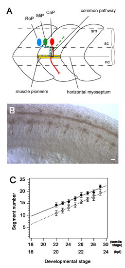
CaP axons extend sequentially starting with those from anterior segments. (A) Schematic representation of the three primary motoneurons (CaP, MiP and RoP) showing their axonal trajectories. CaP axons are projected first to establish the common pathway. The common pathway ends at the muscle pioneers located at the horizontal myoseptum that divides the myotome into dorsal and ventral halves. This point is a choice point where the three axons diverge and follow cell specific pathways to innervate the ventral (CaP), dorsal (MiP) and horizontal myoseptal (RoP) myotomes. sc, spinal cord; nc, notochord; sm, somite. (B) Side view of the trunk of a 28-somite stage (23 hpf) embryos showing that CaP axons labeled with mAb znp1 are more developed anteriorly. Scale bar: 20 �m. (C) The compilation of the stage of initial CaP axon projection (black circle) and the stage when they arrive at the horizontal myoseptum (white circle) by CaPs located in specific segments. CaP axons were projected earlier in anterior segments, e.g. stage 22 for CaPs in segment 14 and stage 29 for those in segment 22. Most CaP axons reached the horizontal myoseptum 1.5 hours after time of initial projection. Each data point represents the mean of at least seven CaPs that were assayed from embryos at 22- to 29-somite stages (20-23.5 hpf). Bars in indicate standard deviations. |