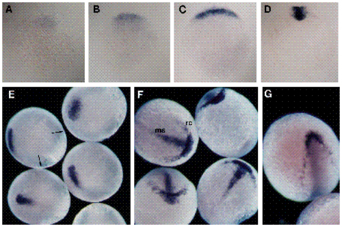
Spatial localization of goosecoid transcripts during embryogenesis. Developmental times are +/- 15 minutes. The blue staining corresponds to goosecoid hybridization. For A-D, the yolks were removed and the blastoderms were photographed in bright field. (A) Animal pole view of ‘early’ 4 h embryo. Expression is limited to a faint patch of cells. (B) Animal pole view of ‘late’ 4 h embryo. The expression domain has expanded to describe a 90° sector. (C) Animal pole view of 5 h embryo with staining limited to cells along the margin. Most cells within the expression domain exhibit staining by this stage. (D) Vegetal pole view of 6 h embryo. Hybridization is restricted to involuted cells within the shield. For E-G, the yolks were left intact, and the embryos were photographed in dark field. (E) Lateral and dorsal views of 8 h embryos. The arrows in the upper left embryo indicate the blastopore at the germ-ring/yolk margin. (F) Anterior and dorsal views of 10 h embryos. The blastopore has closed just ventral to the vegetal pole and the anterior edge of the hybridization is at the animal pole. The medial-strip (ms) and rostral crescent (rc) patterns are indicated in the upper left embryo. The rostral crescent is seen to occupy a position superficial to that of the medial-strip in the top embryo. Staining is sporadic in the lateral regions of the wing-like extensions of the rostral crescent. (G) Dorsal view of 12 h embryo, with sporadic staining along its anterolateral margins. Medial staining is no longer seen.
|
