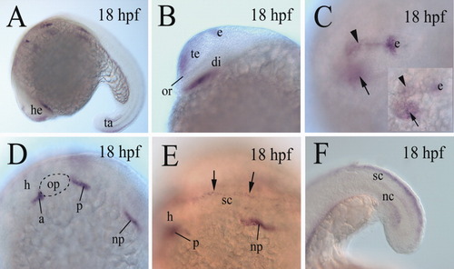
cadherin-6 expression in 18 hours postfertilization (hpf) embryos. A,B: B is a higher magnification image of the head region of the embryo in A, with anterior to the left and dorsal up. C: A higher magnification image of a dorsolateral view of the forebrain (anterior to the left), focusing on the cadherin-6 expression domain (arrowhead) in the dorsal forebrain, whereas the inset shows the same brain, focusing on cadherin-6 expression domain (arrow) in the ventral diencephalon. D: A higher magnification of the hindbrain region (anterior to the left and dorsal up) of the embryo in A. E: A higher magnification of a dorsolateral view of the anterior spinal cord (sc) region (with anterior to the left) of the embryo in A. The two arrows indicate cadherin-6 expression in the dorsal spinal cord. F: A lateral view (anterior to the left and dorsal up) of the tail region of an embryo showing cadherin-6 expression in the dorsal spinal cord. The otic placode in D is outlined with dashed lines. di, diencephalon; e, epiphysis; h, hindbrain; nc, notochord; or, optic recess. The remaining abbreviations are the same as in Figure 1.
|
