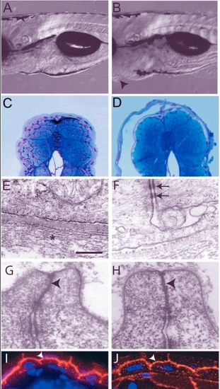
Analysis of the pen mutant phenotype. (A,B) DIC images of 5-day-old wild type (A) and pen mutant larvae (B). The pen mutant larvae exhibit small ventral epidermal growths (arrowheads in B). (C,D) Giemsa stained thin sections of 5-day-old wild-type and mutant larvae. Detachment of epidermis is observed in mutant larvae (D). (E-H) Electron microscopic analysis of the epidermis. Hemidesmosomes are absent and collagen lamella is non-coherent (star) but the basal lamina in between appears normal in the epidermis of pen mutant larvae (E; also compare with Fig. 1E). Desmosomes (arrows in F), however, are present in the mutant epidermis. Tight junctions in the periderm of wild-type and pen mutant (arrowheads in G and H, respectively) show no ultra-structural differences. (I,J) Immunostaining with anti-�-catenin antibody to mark adherens junctions. In wild type (I) as well as in mutant (J), � catenin (arrowheads) is localised at the apical and lateral borders of basal epidermal cells. Staining intensity is lower in the pen mutant. Scale bar: 0.3 mm in A,B; 160 �m in C,D; 543 nm in E; 405 nm in F; 297 nm in G,H; 30 �m in I,J.
|
