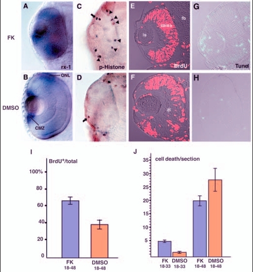
PKA inhibits cell-cycle exit of retinal progenitor cells. (A,B) rx1 expression in 48-hpf forskolin-treated (A) and DMSO-treated (B) retinas. rx1 is expressed both in the outer nuclear layer (ONL) and in the CMZ of DMSO-treated retinas (B); expression is not downregulated in the presence of forskolin (A). (C,D) Labelling of 33-hpf retinas with anti-phosphorylated histone H3 antibody. In the forskolin-treated retina (C), antibody staining was observed in the CMZ (arrow) and at the ventricular surface of the neural retina (arrowheads), whereas only a few mitotic cells were observed in the CMZ (arrow) of the DMSO-treated retina (D). (E,F) BrdU labelling of 48-hpf forskolin-treated (E) and DMSO-treated (F) retinas. In the forskolin-treated retina, most of retinal cells are BrdU positive, whereas cell division seems to cease in the RGC and amacrine cell layers (asterisk) in the DMSO-treated retina. (G,H) TUNEL analysis of 33-hpf forskolin-treated (G) and DMSO-treated (H) retinas. Patterns of cell apoptosis (green) do not correlate with those of neuronal production in forskolin-treated retinas (G; data not shown), although the number of apoptotic cells is higher than in DMSO-treated retinas (H). (I) Ratios of BrdU-positive areas to total area in forskolin- and DMSO-treated retinas. Bars indicate the average ratios that were observed in cryosections of the retinas labelled with the anti-BrdU antibody. The numbers of embryos examined were n=3 and n=2 for forskolin treatment (FK 18-48) and DMSO treatment (DMSO 18-48) between 18 and 48 hpf, respectively. Four sections from the nasal to temporal regions were examined per embryo. (J) Quantification of apoptotic cells in forskolin- and DMSO-treated retinas. Bars indicate the average number of apoptotic cells observed in cryosections corresponding to the central retina subjected to TUNEL. One section was examined per embryo. The numbers of embryos examined were n=7 for forskolin treatment between 18 and 33 hpf (FK 18-33), n=3 for DMSO treatment between 18 and 33 hpf (DMSO 18-33), n=5 for forskolin between 18 and 48 hpf (FK 18-48), and n=3 for DMSO treatment between 18 and 48 hpf (DMSO 18-48). CMZ, ciliary marginal zone; fb, forebrain; FK, forskolin; ls, lens; ONL, outer nuclear layer.
|

