Fig. 2
- ID
- ZDB-FIG-050314-7
- Publication
- Crosnier et al., 2005 - Delta-Notch signalling controls commitment to a secretory fate in the zebrafish intestine
- Other Figures
- All Figure Page
- Back to All Figure Page
|
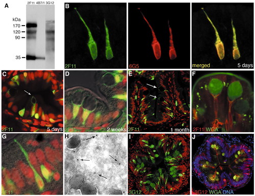
Monoclonal antibodies that mark secretory cells. (A) Western blot of zebrafish gut lysate resolved under non-reducing conditions on SDS-PAGE; antibodies 2F11, 4B7/1 and 3G12 have different antigen specificities. (B) 2F11 (green) and 6G5 (red) stain identical differentiated cells in the intestinal bulb of a 5-day-old larva. (C-H) 2F11 immunolabelling. At 5 days (C), the posterior intestine contains scattered 2F11-positive cells, including a few goblet cells (arrow); goblet cells are more mature and plentiful at 2 weeks (D); by 1 month, goblet cells are also seen in the intestinal bulb (E, arrows). (F) Goblet cells in the intestinal bulb, labelled with wheat-germ agglutinin (green), which stains the mucus compartment, and 2F11 (red), which stains the rest of the cytoplasm. (G) 2F11 (green) also stains some small basal cells and cells that extend thin processes towards the gut lumen; these may be varieties of enteroendocrine cells. (H) Immunogold labelling with 2F11 (small electron-dense particles, arrows) marks the cytoplasm of an enterendocrine cell containing secretory granules (large dark vesicles). (I) 3G12 (green) stains goblet cells in the posterior intestine of a 1-month zebrafish. (J) 3G12 (red) and wheat-germ agglutinin (green) co-stain goblet cells; 3G12 stains both the mucus compartment and the rest of the cytoplasm. Nuclei are stained with TOPRO-3, shown in red (C,D,E,G,I) or blue (J). |

