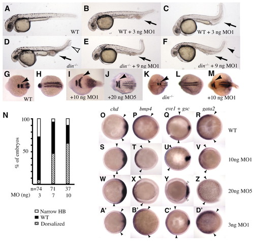
Tsg enhances BMP signaling and can act independently of Chordin. Wild-type or din-/- embryos were injected with the indicated amounts of tsg1-MO1, and photographed on the second day following injection (A-F) or fixed and processed for in situ hybridization (G-M). Some wild-type embryos injected with 3-4 ng of tsg1-MO1 (B,C) displayed a smaller brain and increased blood in the ventral tail (arrows). However, none displayed multiplication of the ventral fin fold, a prominent feature in ventralized din mutants (unfilled arrowhead in D). (E,F) Injection of tsg1-MO1 ameliorated some aspects of the din mutant phenotype; the excess development of blood was reduced (compare arrows in D-F), and in some embryos the multiplication of the fin fold was corrected (black arrowhead in F). However, anterior nervous system development was not rescued. All embryos in A-F are shown from the side, with anterior towards the left. (G-M) Embryos were injected with the indicated amounts of tsg1-MO1 or MO5, fixed at the 8- to 12-somite stage, and processed for in situ hybridization. (I,J) In wild-type embryos, we observed features of dorsalization: lateral expansion of krox20 expression (arrowhead) and myod expression. (L,M) Injection of tsg1-MO1 also dorsalized din-/- embryos. (N) Wild-type embryos injected with the indicated amounts of tsg1-MO1 were fixed and processed for in situ hybridization as above. They were sorted as having a narrowed krox20 expression domain (Narrow HB); as wild type; or as having a widened expression domain for both markers (Dorsalized). The number of embryos is beneath each bar. (O-D′) Wild-type embryos were injected with indicated amounts of tsg1-MOs, fixed at mid-gastrulation (80% epiboly) and processed by in situ hybridization for markers of DV patterning. Expression of chordin, a marker for dorsal ectoderm and mesoderm, was expanded in embryos receiving higher amounts of tsg1-MOs (S,W), and unchanged in embryos receiving a lower amount of MO1 (A′). By contrast, three different markers of ventral territories (bmp4, eve1 and gata2) were decreased (T-V,X,Z) or absent (there is a lack of eve1 in Y) in embryos receiving high amounts of either MO. In the most strongly dorsalized embryos, gsc expression, which marks the dorsal midline tissue, was expanded (unfilled arrowheads in Y). In embryos injected with 3 ng of tsg1-MO1, there was increased expression of gata2, a marker for ventral ectoderm and hematopoetic cells in the ventral mesoderm (D′); expression of the other markers of ventral territories were unchanged (B′,C′). Embryos in G-M are in dorsal view, with anterior towards the left. (G,H and K,L) Two views of a single embryo rotated to show the krox20 or myod expression. All embryos in O-D′ are shown from the animal pole, with dorsal towards the right; arrowheads indicate the lateral limits of dorsal or ventral markers.
|

