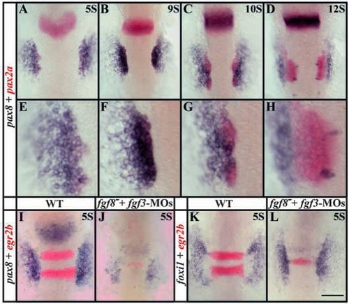
pax8 expression partially overlaps with pax2a in the preotic region and, like pax2a, depends upon Fgf signaling. Expression domains of pax8, (blue) and pax2a (red) coincide in the preotic region (A-C) but diverge after formation of the placode (D). At five-somite (A,E) to nine-somite (B,F) stages, the pax8 domain encompasses pax2a but also extends further laterally. By the 10- somite stage, when the otic placode is forming, pax8 expression diminishes in the placode (C,G) and is completely absent from the placode by the 12-somite stage (D,H) but is obvious just lateral to the placode. The bilateral pairs of medial blue spots in D,H are hindbrain neurons that express pax8. (E-H) High-magnification views of embryos shown in A-D. (I,J) Fgf3 and Fgf8 depletion leads to a severe reduction of pax8 expression at the five-somite stage but not to complete loss. In Fgf3- and Fgf8-depleted embryos, only weak expression can be detected (J) compared with uninjected wild-type embryos (I). Expression of egr2b (red) in the hindbrain (I) is also reduced when Fgf signals are lost (J) (Maves et al., 2002). (K,L) foxi1 is not dependent on the Fgf3 and Fgf8 signal. In wildtype embryos at the five-somite stage, foxi1 is expressed in two patches lateral to the neural plate (K) and depletion of Fgf3 and Fgf8 has no obvious effect on foxi1 expression (L). Dorsal views, anterior towards the top. Scale bar: 120 μm for A-D,I-L; 40 μm for E-H.
|
