
Fig. 4
- ID
- ZDB-FIG-240702-134
- Publication
- Meneghetti et al., 2020 - Zebrafish ambra1a and ambra1b Silencing Affect Heart Development
- Other Figures
- All Figure Page
- Back to All Figure Page
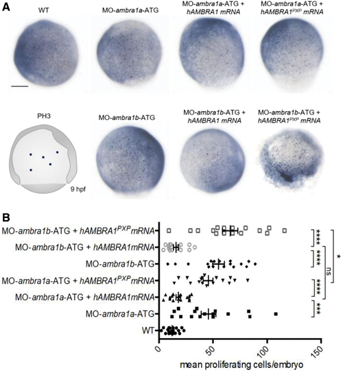
AMBRA1 morphant embryos display increased cell proliferation. (A) Representative images of phospho-histone H3 immunohistochemistry in WT and AMBRA1 morphant embryos at 9 hpf. Scale bar, 200 μm. (B) The numbers of proliferating cells were counted and compared between WT embryos, AMBRA1 morphant embryos, and AMBRA1 morphant embryos co-injected with hAMBRA1 or mutated hAMBRA1PXP mRNAs. For each condition, 10 embryos were analyzed, and the experiment was performed twice. Statistical analysis was performed using Student's t-test. *p < 0.05; ***p < 0.001; ****p < 0.0001. Color images are available online. |