
FIGURE
Fig. 4
- ID
- ZDB-FIG-230829-22
- Publication
- George et al., 2022 - Zebrafish model of RERE syndrome recapitulates key ophthalmic defects that are rescued by small molecule inhibitor of shh signaling
- Other Figures
- All Figure Page
- Back to All Figure Page
Fig. 4
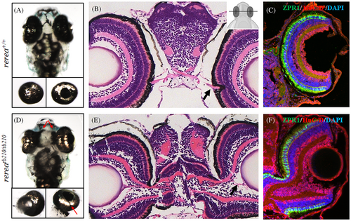
Zebrafish rerea homozygous mutants exhibit disruption of retinal optic stalk boundary. Zebrafish larvae homozygous for rerea mutation exhibit disruption of retinal optic stalk boundary (A–C, D–F), which was further confirmed by isolation of eye cups (A, D, bottom panels) and detailed coronal histological sectioning (B, E) of 6 dpf larvae. Unfused margins of optic fissure can be observed (D bottom panel, red arrow). The presence of ectopic differentiated retinal layers (C, F) was confirmed by immunostaining with ZPR-1 (photoreceptors) and HuC/D (amacrine cells and ganglion cells).
|
Expression Data
| Antibodies: | |
|---|---|
| Fish: | |
| Anatomical Terms: | |
| Stage: | Day 6 |
Expression Detail
Antibody Labeling
Phenotype Data
| Fish: | |
|---|---|
| Observed In: | |
| Stage: | Day 6 |
Phenotype Detail
Acknowledgments
This image is the copyrighted work of the attributed author or publisher, and
ZFIN has permission only to display this image to its users.
Additional permissions should be obtained from the applicable author or publisher of the image.
Full text @ Dev. Dyn.