
FIGURE
Fig. 3
- ID
- ZDB-FIG-230829-21
- Publication
- George et al., 2022 - Zebrafish model of RERE syndrome recapitulates key ophthalmic defects that are rescued by small molecule inhibitor of shh signaling
- Other Figures
- All Figure Page
- Back to All Figure Page
Fig. 3
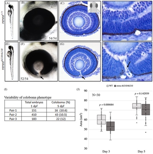
Zebrafish rerea homozygous mutants exhibit coloboma. Three-day post fertilization WT zebrafish embryos (A, B) exhibit fused optic fissure margins (54 genotyped embryos), whereas rerea homozygous mutants exhibit coloboma (E, F, 52/54 genotyped embryos). Sagittal histological sectioning of WT (C, D, N = 10 genotyped embryos) and rerea mutant (G, H, N = 10 genotyped embryos) zebrafish eye (3 dpf). The schematic in C gives the approximate plane of sectioning for the histologic sections. Coloboma showed variable penetrance in embryos at 5 dpf (I). Measurement of eye area (lateral view, Figure 2K) revealed significantly smaller eye at 3 dpf (P < .05) of rerea mutant larvae compared with WT. No statistical difference in eye size was observed at 5 dpf (J). Student's t test was used to determine level of significance and P value is provided (N = 30 genotyped embryos/group).
|
Expression Data
Expression Detail
Antibody Labeling
Phenotype Data
| Fish: | |
|---|---|
| Observed In: | |
| Stage Range: | Protruding-mouth to Day 5 |
Phenotype Detail
Acknowledgments
This image is the copyrighted work of the attributed author or publisher, and
ZFIN has permission only to display this image to its users.
Additional permissions should be obtained from the applicable author or publisher of the image.
Full text @ Dev. Dyn.