
FIGURE
Fig. 1
- ID
- ZDB-FIG-230829-19
- Publication
- George et al., 2022 - Zebrafish model of RERE syndrome recapitulates key ophthalmic defects that are rescued by small molecule inhibitor of shh signaling
- Other Figures
- All Figure Page
- Back to All Figure Page
Fig. 1
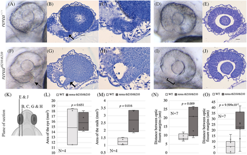
Zebrafish rerea homozygous mutants exhibit enlarged optic stalk and optic fissure margins that are not apposed. Brightfield images of live fish embryos (A, F), ventro-lateral angle focused on the optic stalk [OS] and histology sections of wild-type (B, C) and rerea mutant (G, H) zebrafish embryos (24 hpf) where OS (black arrow) is present on the ventral side of the eye (lateral view). Brightfield images (D, I) and histology sections of wild-type (E) and rerea mutant (J) zebrafish embryos (24 hpf) depicting optic fissure margins that are far apart (square bracket) on the ventral side of the eye (lateral view). Schematic of plane of histology sections (K). Quantification of area of the eye (L) and optic stalk (M). Distance between optic fissure margins of brightfield images (N) and histology images (O) displayed as a box and whisker plot with dots indicating individual measurements and the horizontal bar the median value. Student's t-test was applied for P values. Scale bar is 20 μm.
|
Expression Data
Expression Detail
Antibody Labeling
Phenotype Data
| Fish: | |
|---|---|
| Observed In: | |
| Stage: | Prim-5 |
Phenotype Detail
Acknowledgments
This image is the copyrighted work of the attributed author or publisher, and
ZFIN has permission only to display this image to its users.
Additional permissions should be obtained from the applicable author or publisher of the image.
Full text @ Dev. Dyn.