
- Title
-
Involvement of Glucosamine 6 Phosphate Isomerase 2 (GNPDA2) Overproduction in β-Amyloid- and Tau P301L-Driven Pathomechanisms
- Authors
- Lachťn-Montes, M., Cartas-Cejudo, P., Cortťs, A., Anaya-Cubero, E., Peral, E., AusŪn, K., DŪaz-PeŮa, R., FernŠndez-Irigoyen, J., SantamarŪa, E.
- Source
- Full text @ Biomolecules
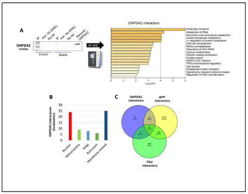
Identification of GNPDA2 molecular interactors. ( |

RNA-seq analysis in hNECs. ( |

Gene interactome maps for differentially expressed genes in hNECs. Representation of the interactions between differentially expressed genes after GNPDA2 overexpression ( |
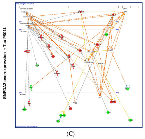
Gene interactome maps for differentially expressed genes in hNECs. Representation of the interactions between differentially expressed genes after GNPDA2 overexpression ( |
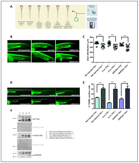
Study of GNPDA2 overexpression in zf Tau P301L transgenic embryos. ( |

Proteomics in h.Tau P301L zebrafish embryos. ( |
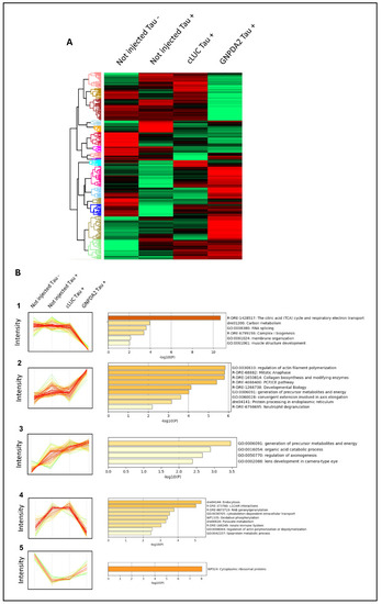
Differentially expressed proteins across Tau P301L embryos overexpressing GNPDA2. ( |

|