FIGURE SUMMARY
Title

Lymphatic Defects in Zebrafish sox18 Mutants Are Exacerbated by Perturbed VEGFC Signaling, While Masked by Elevated sox7 Expression

Authors
Moleri, S., Mercurio, S., Pezzotta, A., D'Angelo, D., Brix, A., Plebani, A., Lini, G., Di Fuorti, M., Beltrame, M.
Source
Full text @ Cells

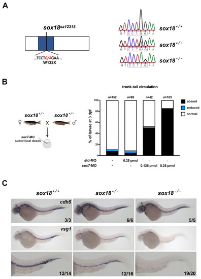
The sox18sa12315 mutant behaves as expected for a null allele. (A) On the left is a schematic representation of the Sox18 protein, with the HMG-box domain in blue. The G > A transition and the premature stop codon introduced in the sa12315 mutant are indicated. Fragments of the electropherograms derived using Sanger sequencing of the region surrounding the mutation in wt (sox18+/+), heterozygous (sox18+/−) or homozygous mutants (sox18−/−) are reported on the right. The restriction site for the BstNI/MvaI enzymes (boxed sequence) is disrupted by the mutation. (B) Embryos derived from sa12315 heterozygote matings and injected with subcritical doses of sox7-MO were collected in several independent experiments and analyzed in vivo at 2 dpf and 3 dpf or fixed at around 30 hpf for ISH, as shown in (C). The histogram on the right shows the trunk–tail circulatory phenotypes observed at 3 dpf. In control embryos, i.e., uninjected or injected with a standard control MO (first and second bars, respectively), trunk–tail circulatory defects are present in a small percentage of embryos. On the contrary, the partial knockdown of sox7 causes a blockage in trunk–tail circulation in a dose-dependent manner (third and fourth bars). Circulatory defects are genotype-dependent (see Table S1). (C) ISHs were performed on embryos derived from sa12315 heterozygote matings and injected with subcritical doses of sox7-MO, as shown in (B), fixed at around 30 hpf. Upper panels show control ISH performed with the endothelial marker cdh5, showing no gross alteration in embryos of the three different genotypes. Lower panels show ISHs performed with a probe for vsg1/plvapb, whose expression was particularly downregulated in double partial sox7/sox18 morphants [29]. Higher magnification images of the trunk–tail regions of the embryos are also shown. Experiments were repeated twice; all plavpb stained embryos and a subset of cdh5 stained embryos were genotyped; numbers in each image refer to a single experiment. Lateral views, anterior to the left. Pictures were taken at 40× and 63× magnification, for lower and higher magnification images respectively.

EXPRESSION / LABELING:
Genes:
Fish:
Knockdown Reagent:
Anatomical Term:
Stage: Prim-15
PHENOTYPE:
Fish:
Knockdown Reagent:
Observed In:
Stage Range: Prim-15 to Protruding-mouth

Homozygous sox18 mutants show subtle but statistically significant defects in thoracic duct (TD) formation. (A) Confocal trunk images representing wt (+/+) and sox18sa12315 homozygous mutant larvae (−/−) in the Tg(lyve1b:DsRed) line at 5dpf. Large and small white arrowheads point to TD+ segments (of typical or thinner aspect, respectively) while asterisks indicate the absence of TD. (B) The graph reports the mean number of TD+ segments, counted along 10 consecutive trunk segments, together with the Standard Error of the Mean (SEM), in all analyzed embryos of the three genotypes (wt: sox18+/+, het: sox18+/−, hom: sox18−/−). Data were gathered in three independent experiments, and each symbol represents the number of TD+ segments of a single analyzed larva. n = number of larvae, ** = p < 0.01. TD = thoracic duct; DA = dorsal aorta; PCV = posterior cardinal vein. Lateral view, anterior to the left.

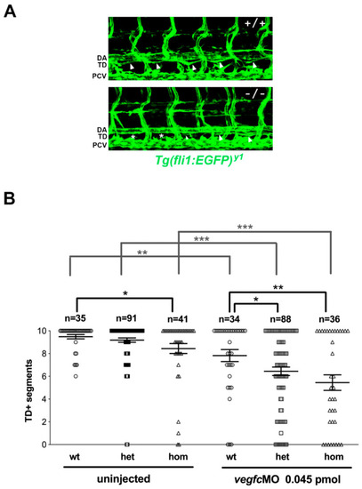
TD formation defects are exacerbated upon slight perturbation of Vegfc signaling. The progeny of sox18sa12315 heterozygote matings in the Tg(fli1a:EGFP)y1 line were injected with a subcritical dose of vegfc-MO or left uninjected; TD formation was analyzed at 5dpf. (A) Confocal trunk images of uninjected wt (+/+) and sox18 homozygous mutant (−/−) larvae. Arrowheads point to TD+ segments, while asterisks indicate the absence of TD; a smaller arrowhead marks a thinner TD+ segment. (B) The graph reports the mean number of TD+ segments, counted along 10 consecutive trunk segments, together with the SEM, for all analyzed larvae of each genotype (wt: sox18+/+, het: sox18+/−, hom: sox18−/−). Each symbol represents the number of TD+ segments of a single larva; data were gathered in several independent experiments. Uninjected larvae on the left are compared to larvae with partially reduced Vegfc on the right. n = number of larvae, * = p < 0.05; ** = p < 0.01; *** = p < 0.001. TD = thoracic duct; DA = dorsal aorta; PCV = posterior cardinal vein. Lateral view, anterior to the left.

The expression of sox7 in the PCV is upregulated in sox18 mutants, but not in sox18 morphants. (A) Representative images of sox7 ISH on embryos at around 26 hpf derived from matings of sox18sa12315 heterozygotes in the Tg(lyve1b:DsRed) line. Higher magnifications of the trunk region are shown below the full size images. Compared to wt embryos, the sox7 ISH signal in the PCV is elevated in the great majority of sox18−/− homozygotes and, to a lesser extent, in sox18+/− heterozygotes. Numbers in each image state the number of embryos with the reported phenotype over the total analyzed embryos in one representative experiment. Lateral views, anterior to the left. Pictures were taken at 40× and 63× magnification, for lower and higher magnification images respectively. (B) Left, representative ImageJ-modified images, used to perform the quantification of the sox7 ISH signal in the PCV and the DA (as described in Materials and Methods) on 26 hpf wt (+/+), sox18sa12315 heterozygotes (+/−) or homozygous mutants (−/−). The graph on the right shows the calculated PCV/DA ratio in each embryo; embryos are grouped based on their genotypes: mean values and SEM are indicated. (C) The same analysis was performed on sox7 ISH of sox18 morphants and control embryos, as shown in Figure S6. The calculated PCV/DA ratio of each embryo is shown in the graph; mean values and SEM for std-MO injected embryos and sox18 morphants are indicated. n = number of embryos, ** = p < 0.01; DA = dorsal aorta; PCV = posterior cardinal vein. The analysis was also repeated on ISHs of sox18sa12315 mutants in the Tg(fli1a:EGFP)y1 reporter line with similar results. ISH experiments on sox18sa12315 mutants were repeated at least three times. Data shown in A and B were generated on different clutches of embryos.

ZFIN is incorporating published figure images and captions as part of an ongoing project. Figures from some publications have not yet been curated, or are not available for display because of copyright restrictions.

ZFIN is incorporating published figure images and captions as part of an ongoing project. Figures from some publications have not yet been curated, or are not available for display because of copyright restrictions.

ZFIN is incorporating published figure images and captions as part of an ongoing project. Figures from some publications have not yet been curated, or are not available for display because of copyright restrictions.

ZFIN is incorporating published figure images and captions as part of an ongoing project. Figures from some publications have not yet been curated, or are not available for display because of copyright restrictions.

EXPRESSION / LABELING:
Gene:
Fish:
Knockdown Reagent:
Anatomical Terms:
Stage: Prim-5
Acknowledgments
This image is the copyrighted work of the attributed author or publisher, and ZFIN has permission only to display this image to its users. Additional permissions should be obtained from the applicable author or publisher of the image. Full text @ Cells