FIGURE SUMMARY
Title

Real-time monitoring of endogenous Fgf8a gradient attests to its role as a morphogen during zebrafish gastrulation

Authors
Harish, R.K., Gupta, M., Zöller, D., Hartmann, H., Gheisari, A., Machate, A., Hans, S., Brand, M.
Source
Full text @ Development

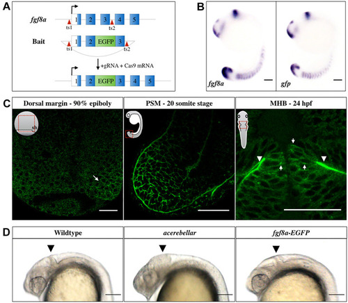
Generation and characterization of the Tg(fgf8a:fgf8a-EGFP) fish line. (A) Knock-in strategy: EGFP was inserted between exons 2 and 3 of the endogenous Fgf8a locus using CRISPR/Cas9. Exon sequences are shown as numbered blocks, separated by introns. The open reading frame sequence is in blue. Red arrowheads indicate the sgRNA target sites (ts). (B) In situ hybridization against fgf8a and gfp in 16-somite stage (ss) embryos. Scale bars: 100 μm. (C) Fgf8a-EGFP fluorescence in the dorsal embryonic margin (arrow) at late gastrula, PSM at 20 ss, as well as at the basal (arrowheads) and lateral (arrows) sides of the MHB neuroepithelia 24 h post fertilization (hpf). Orientations of embryos are shown schematically in insets. Red rectangles outline the locations imaged. sh, shield. Scale bars: 50 μm. (D) Comparison of homozygous fgf8a-EGFP with wild type and homozygous ace mutants at 24 hpf. Homozygous viability and normal structure of the MHB (arrowhead) in the fgf8a-EGFP transgenics confirms the functionality of the fgf8a-EGFP knock-in line. Lateral views are shown. Scale bars: 100 μm.

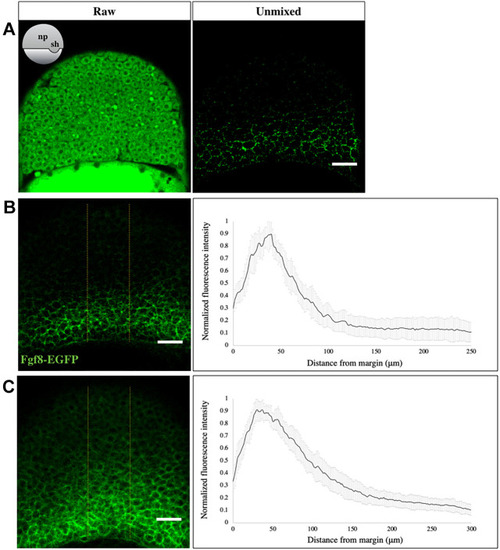
Fgf8a-EGFP forms a gradient during gastrulation. (A) EGFP fluorescence in an optical section of the neural plate in early gastrula stage fgf8a-EGFP embryos, as visualized using the GaAsP detector, before (left) and after (right) linear unmixing. The orientation of embryos is shown schematically in the inset. np, neural plate; sh, shield. (B,C) Sum-intensity z-projected images, derived after linear unmixing (left), used to extract fluorescence intensity profiles in the neural plate (yellow boundaries) at early (B) and mid-gastrula (C) stages. The analysis reveals a graded distribution of Fgf8a towards the animal pole (right). N=15 embryos. Scale bars: 50 μm. Data are mean±s.d.

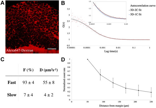
Fgf8a-EGFP propagates by diffusing via the extracellular space. (A) Extracellular space (ECS) in an early gastrula embryo, as visualized by Alexa-647-tagged Dextran injection. The orientation of the embryo is as in Fig. 2A. Scale bar: 50 μm. (B) FCS autocorrelation curve from the ECS of a Tg(fgf8a:fgf8a-EGFP) embryo fitted with 2-C (black curve) and 1-C (blue curve in inset) 3D-diffusion models. (C) Results from fitting with the 3D-2C diffusion model. F, fraction of each component; D, diffusion coefficient. n=52 measurements. (D) Plot of count rate, derived from FCS analysis versus distance from the margin at early gastrula showing graded distribution of Fgf8a in the ECS along the animal-vegetal axis. N=20 embryos. Data are mean±s.d.

Input-output relationship of Fgf8a-mediated patterning during gastrulation. (A) smFISH against tbxta and etv4 in early-gastrula stage embryos, after threshold adjustment for background subtraction. (B) smFISH against etv4 in DMSO- (left) and SU5402- (right) treated embryos at early gastrula stage. In both A and B, optical sections of lateral views are shown. Scale bars: 20 μm. (C) Maximum-intensity z-projected images for smFISH against tbxta (top) and etv4 (bottom) at early gastrula (left), and plot of normalized transcript number versus distance from the margin (right). N=13 embryos for tbxta, N=7 for etv4. For A-C, DAPI (blue) was used as a nuclear marker. Overlay with the Fgf8a distribution profile (orange curve) determines its relative extracellular levels corresponding to maximal expression domains of both the transcripts (see blue lines). Peak of Fgf8a-EGFP profile corresponds to an absolute concentration of ∼8 nM. Scale bars: 50 μm. (D) Venus fluorescence in DMSO- (left) and SU5402- (right) treated Tg(etv5b:etv5b-Venus) embryos at early gastrulation. Optical sections of laterally mounted embryos are shown. Scale bars: 50 μm. (E) Sum-intensity z-projected image of Etv5b-Venus from early gastrula-staged transgenic embryos (left) and analysis of nuclear fluorescence intensity as a function of distance from the margin (right). Overlay with the Fgf8a gradient curve (orange curve) shows its minimum relative abundance in the extracellular spaces that correspond to the peak Etv5b output (see blue lines). N=11 embryos. Scale bars: 50 μm. For C and E, exact orientations are shown schematically in insets. Data are mean±s.d.

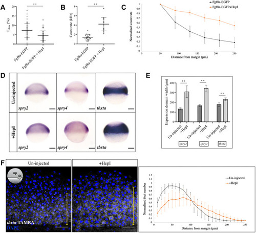
Manipulation of the Fgf8a input alters the signaling output. (A) Injection of HepI reduces the proportion of the slow-moving component of Fgf8a-EGFP, as detected by FCS, in the extracellular spaces. n=52 measurements for Fgf8a-EGFP, n=56 for HepI-injected. **P<0.0001. (B) FCS count rates for Fgf8a-EGFP in the extracellular spaces near the embryonic margin in uninjected and HepI-injected embryos. Count rate is increased upon HepI injection. n=20 for uninjected, n=17 for HepI. **P<0.0001. (C) Plot of count rate versus distance from margin in control (black) and HepI-injected (orange) cases. N=20 embryos for control, N=13 for HepI. (D) In situ hybridization against spry2, spry4 and tbxta in control and HepI-injected early gastrula embryos. Lateral views are shown. Scale bars: 100 μm. (E) Quantification of expression domains of genes as in D, along the animal-vegetal axis. **P<0.0001. (F) smFISH against tbxta in uninjected and HepI-injected embryos at early gastrula, and plot of normalized transcript number versus distance from the margin. tbxta expression is reduced near the margin and increased in its range away from the margin (yellow line) upon HepI injection. Orientation of embryos is shown schematically in inset. N=10. Scale bars: 50 μm. Data are mean±s.d.

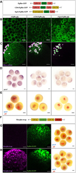
Extracellular diffusion of Fgf8a is necessary for its morphogenic activity. (A) The three different versions of Fgf8a were used in this study. sp, signal peptide; TM, transmembrane. (B) Confocal images of late blastula-stage embryos injected with the various constructs at the one-cell stage to assess localization. Scale bars: 20 μm. (C) Generation of ectopic clones of the constructs and imaging at late blastula stages to determine protein spreading. Co-injection with HRas-mKate labels the source cells (magenta). Scale bars: 50 μm. (D) In situ hybridization against spry4 at late blastula after generating ectopic clones as in C. (E) Double in situ hybridization against gfp (red) and spry4 (blue) in such embryos at late blastula to depict the ectopic source and target gene induction domains, respectively. Scale bars: 100 μm. (F) Schematic of the Morpho-trap construct. (G,H) Late blastula-stage embryos injected at the 32-cell stage with Fgf8a-EGFP (G) or the Morpho-trap at the one-cell stage followed by Fgf8a-EGFP at the 32-cell stage (H). Confocal images are shown in the first two panels. Scale bars: 50 μm. The third panel depicts double in situ hybridization against gfp (red) and spry4 (blue) in such embryos to indicate the Fgf8a-EGFP clone and target gene induction domain, respectively. Scale bars: 100 μm. Animal pole views of embryos are shown throughout.

Schematic summary of the work. (A) Visualizing endogenous Fgf8a via knock-in reveals its graded distribution during gastrulation (left). Single-molecule FCS detects Fgf8a as an extracellularly diffusing morphogen (enlarged on the right). HS, heparan sulfate. (B) Membrane tethering of Fgf8a in cell clones restricts its signaling range.

Acknowledgments
This image is the copyrighted work of the attributed author or publisher, and ZFIN has permission only to display this image to its users. Additional permissions should be obtained from the applicable author or publisher of the image. Full text @ Development